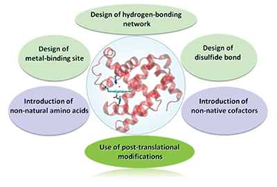
Loading...
中文
Announcement
More
Progress in Chemistry 2018, No.10 Previous issue Next issue

In this issue:

Review
Rational Design of Artificial Metalloenzymes: Case Studies in Myoglobin
Yingwu Lin
2018, 30 (10): 1464-1474 | DOI: 10.7536/PC180528
Published: 15 October 2018
Abstract
Metalloenzymes play diverse important functions in biological systems. Meanwhile, design of artificial metalloenzymes may fine-tune and expand the functionalities of natural metalloenzymes, and even create novel enzymes with more advanced functions. Myoglobin(Mb) is an ideal protein model for design of heme proteins and other metalloenzymes. In recent years, various approaches have been developed for design of artificial metalloenzymes based on the protein scaffold of Mb, which include design of hydrogen-bonding network, intramolecular disulfide bond, the use of post-translational modifications, introduction of non-natural amino acids and non-native cofactors. This review summaries the recent progress in these aspects, which will help us understand the structure and function relationship of metalloenzymes, master the idea and methodologies of artificial metalloenzyme design, thereby promoting the rapid development in this field.
Contents
1 Introduction
2 Approaches and cases studies
2.1 Design of hydrogen-bonding network
2.2 Design of metal-binding site
2.3 Design of disulfide bond
2.4 Use of post-translational modifications
2.5 Introduction of non-natural amino acids
2.6 Introduction of non-native cofactors
3 Conclusion and outlook
SOD1 Inhibition Regulates the ROS Signaling Transduction
Xiang Li, Jiayuan Shi, Shuang Qiu, Mingfang Wang, Changlin Liu*
2018, 30 (10): 1475-1486 | DOI: 10.7536/PC180608
Published: 15 October 2018
Abstract
Copper-zinc superoxide dismutase(SOD1) is an antioxidant metalloenzyme that widely distriutes within a cell, and catalyzes rapid dismutation of superoxide anion(O2·-) into hydrogen peroxide(H2O2) and oxygen(O2) to maintain the homeostasis of intracellular reactive oxygen species(ROS). The growth and proliferation of cancer cells depend on higher concentrations of H2O2, whereas high expression of SOD1 can maintain higher H2O2 levels and ROS homeostasis of cancer cells. SOD1 inhibition effectively regulates ROS signaling pathways in a cancer cell, inhibits cancer cell growth and proliferation, arrests cancer cell cycle, and promotes cancer cell apoptosis. Targeting SOD1 can regulate the ROS signaling network and selectively kill cancer cells. This review summarizes various types of SOD1 inhibitors designed so far, the regulation of ROS signaling transduction by SOD1 inhibition, and the mechanism of specific SOD1 inhibition-mediated apoptosis of cancer cells.
Contents
1 Introduction
2 The structure and function of SOD1
2.1 Structure and catalytic mechanism of SOD1
2.2 Functions of SOD1
3 Regulation of ROS signaling pathways by SOD1
3.1 SOD1 and ROS signaling pathways
3.2 SOD1 and cancer
4 SOD1 inhibition
4.1 Inhibition of SOD1 expression
4.2 SOD1 inhibitors
5 SOD1 inhibition-regulated ROS signaling pathways
6 Conclusion
The Structure and Function of Selenoprotein S and Its Relationship with Diseases
Hongmei Liu*, Jianbo Jin, Jun Zhou, Kaixun Huang, Huibi Xu
2018, 30 (10): 1487-1495 | DOI: 10.7536/PC180613
Published: 15 October 2018
Abstract
Selenium is an essential trace element for human being and its biological functions are mainly carried out by selenoproteins. Selenoprotein S(SELENOS) mainly localizes to the endoplasmic reticulum(ER) membrane and is involved in the process of ER-associated degradation. The biological functions of SELENOS are mainly carried out through the coiled-coil domain and C-terminal disordered structure region containing selenocysteine residue in the cytosol. A large number of in vitro studies have shown that SELENOS participates in the regulation of oxidative stress, ER stress and inflammation, and thus is possibly involved in the development of cardiovascular disease, type 2 diabetes, and Alzheimer's disease. Furthermore, the observational epidemiological studies have found that many single nucleotide polymorphisms in the SELENOS gene are closely associated with cardiovascular disease and cancer. This paper reviews the structure and function of SELENOS and its relationship with diseases. The problems remaining to be solved are summarized and the future developments are prospected.
Contents
1 Introduction
2 Expression pattern, structure, and distribution of SELENOS
3 The biological function of SELENOS
3.1 Regulation of oxidative stress
3.2 Regulation of endoplasmic reticulum stress
3.3 Regulation of inflammation
4 The relationship of SELENOS with diseases
4.1 Cardiovascular diseases
4.2 Type 2 diabetes
4.3 Cancer
4.4 Alzheimer's disease
5 Conclusion and outlook
Selenoprotein R: A Unique Methionine Sulfoxide Reductase
Tengrui Shi, Yujie Yang, Qiong Liu, Nan Li*
2018, 30 (10): 1496-1502 | DOI: 10.7536/PC180625
Published: 15 October 2018
Abstract
Selenoproteins represent a category of proteins that possess selenocysteine as the active center, by taking advantage of robust reducing ability of selenium, selenoproteins exert an important antioxidant function in various organisms. To date, there are 25 genes that encode selenoproteins found in human genome. Among them, selenoprotein R is the only methionine sulfoxide reductase that contains selenocysteine. It is located in the cytoplasm and nucleus, given the protein structure and strong nucleophilicity of selenium, selenoprotein R is competent to specifically reduce the oxidized sulfur in methionine-R-sulfoxide. Selenoprotein R could directly interact with many proteins, such as actin, transient receptor potential channel proteins, and β-amyloid protein. It may play crucial roles in the central nervous system and is closely related with the development of neurodegenerative diseases.
Contents
1 Introduction
2 The recognition of selenoprotein R and other Msrs
3 Phenotype induced by Msrs knockout
4 Involvement of selenoprotein R in neural degenerative disease
5 Conclusion and outlook
Collagen Mineralization and Tissue Repair
Yuanyuan Wu, Haihua Pan, Ruikang Tang
2018, 30 (10): 1503-1510 | DOI: 10.7536/PC180541
Published: 15 October 2018
Abstract
The hard tissue(tooth and bone) in vertebrates is formed by the biomineralization processes, which are regulated and controlled by bio-systems. Among them, the mineralized collagen fibril is an essential building block for the construction of biominerals. The mineralized collagen is a hybrid material of nano inorganic mineral(calcium phosphate) and assembled organic molecules(collagen). The advanced nano hierarchical and ordered structure of the mineralized collagen fibrils will endow biological hard tissue with excellent mechanical properties(such as hardness and toughness). The structure feature and mineralization process may inspire new materials fabrications aimed at hard tissue repair. It is important but challenging to reconstruct the ordered organic/inorganic hybrid structures via intrafibrillar mineralization of collagen, and that is the key to the tissue repair. In this review, the same basic questions are discussed including hierarchical structures of bones, the assembled structure of collagen fibrils and the nano-hybrid structure of mineralized collagen fibrils, the interactions between the organic(collagen and non-collagenous proteins) and inorganic counterparts and their interface, the modification of mineralizing interface by the functional molecules, and the control of collagen mineralization and their applications in tissue repair. Some unresolved but urgent issues for collagen mineralization are also discussed, such as the regulation of amorphous precursors for the infiltration into the nano-compartment of collagen fibrils, the control of the mineralization rate and degree, as well as the large scale fabrication of ordered collagen fibrils and their mineralization.
Contents
1 Introduction
2 Hierarchical organic-inorganic structure of mineralized collagen
3 Interactions between the organic and inorganic components
4 Interfaces of collagen mineralization and their regulation
5 Biomimetic mineralization of collagen fibrils and their applications in hard tissue repair
6 Conclusion
Biomimetic Mineralization Synthesis of Nanomaterials Under the Mediation of Cells and Potential Applications
Xiaoxiao Xie, Xiaoming Ma*, Xiangli Ru, Yi Chang, Yuming Guo, Lin Yang*
2018, 30 (10): 1511-1523 | DOI: 10.7536/PC180807
Published: 15 October 2018
Abstract
Biomineralization has endowed many organisms with peculiar functions under the modification of the inorganic nanostructure biosynthesized under the mediation of the cells. Recently, with the development of biosynthesis and nanotechnology, using organisms to synthesize nanomaterials has attracted the wide attention of researchers. Compared with traditional physical and chemical synthesis methods, biosynthesis has the advantages of simple operation and environmental friendliness. So far, there have been many studies on the use of bacteria, fungi, some plants and human cells to successfully produce various shapes and sizes of inorganic nanomaterials. The obtained nanomaterials not only have good dispersibility and stability, but also have excellent application performance. Hence, it is widely used in the fields of catalysis, electrology, optics and biomedicine, etc. The biomimetic mineralization synthesis of nanoparticles is likely to provide scientific reference for the discovery of novel nanomaterials. This review presents the related knowledge of biological nanotechnology and the research progresseof nanomaterials synthesized by different organisms. The current main problems of biosynthetic nanomaterials, progress of research contents and innovation points of are elaborated in detail.
Contents
1 Introduction
2 Biomimetic mineralization synthesis of nanomaterials under the mediation of living cells
2.1 Bacterial cells
2.2 Fungus cells
2.3 Human cells
2.4 Plant cells extract
3 Study of biosynthetic mechanism of nanomaterials under the mediation of living cells
3.1 Biosynthetic mechanism of nanomaterials under the mediation of living cells
3.2 Regulating factors of biosynthesis mechanism of nanomaterials
4 Applications of the nanomaterials
5 Conclusion and outlook
Real-Time Luminescence Tracking in Living Cells with Metal Complexes
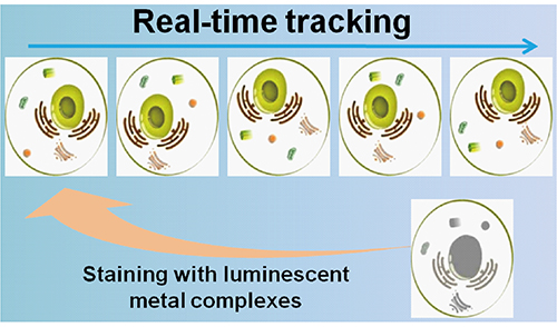
Kangqiang Qiu, Hongyi Zhu, Liangnian Ji, Hui Chao
2018, 30 (10): 1524-1533 | DOI: 10.7536/PC180703
Published: 15 October 2018
Abstract
Metal complexes possess outstanding photophysical properties, such as large Stokes shifts, good photostability, long luminescent lifetimes, two-photon luminescent properties and so on. During the past years, metal complexes have attracted intensive research interest in biological imaging for their excellent properties. Moreover, they also have been applied for real-time tracking the cellular dynamics during a series of biological events. In this review, the essential requirements of the metal complexes for cellular dynamic real-time tracking are summarized. The specificity, the good photostability and the low cytotoxicity of metal complexes are the key factors for real-time tracking. According to the optical properties, the imaging modes (single photon imaging, multiphoton imaging, luminescence lifetime imaging) of the metal complexes for real-time tracking are outlined. Furthermore, the advantages and disadvantages of the imaging modes during cellular real-time dynamic tracking are compared. As for the objects of real-time tracking, the article focuses on the organelles and the microenvironment factors. The real-time dynamic tracking of organelles (mitochondria, lysosomes, lipid droplets) and microenvironment factors (hypoxia, pH, viscosity, temperature, polarity) are detailed overviewed and systemized the related research directions. In the end, some existing problems and the challenges of the metal complexes during real-time tracking are presented and discussed.
Contents
1 Introduction
2 The essential requirements of real-time tracking probes
3 The imaging modes of transition metal complexes
4 Cellular dynamics tracking
4.1 Tracking the dynamics of organelles
4.2 Tracking the dynamics of microenvironment factors
5 Conclusion and outlook
Design and Application of T1-T2 Dual-Modal MRI Contrast Agents
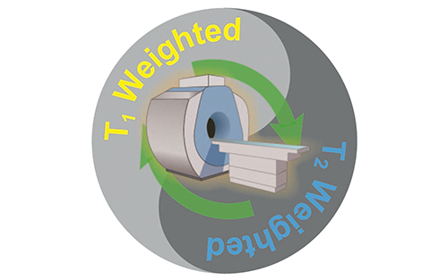
Guang Deng, Hong Yang, Zhiguo Zhou*, Shiping Yang*
2018, 30 (10): 1534-1547 | DOI: 10.7536/PC180537
Published: 15 October 2018
Abstract
Magnetic resonance imaging(MRI) has been used in the clinic widely as an early noninvasive diagnosis technique. According to the theory of MRI, three imaging modes exist, namely, T1-weighted imaging, T2-weighted imaging and diffusion weighted imaging. Accordingly, there are two types of exogenous MRI contrast agents, namely T1 contrast agent and T2 contrast agent, for improvement in contrast in MR images. Either T1 or T2 contrast agents has its own advantages and disadvantages, respectively. Recently, a kind of new contrast agents, T1-T2 dual-modal contrast agent, has been designed, which can be used on one machine with an accurate match of spatial and temporal imaging parameters. This article reviews the progress of design and chemical synthesis methods of T1-T2 dual-mode contrast agents, and also introduces their biomedical applications.
Contents
1 Introduction
2 The research about dual T1-T2 MRI contrast agents
2.1 Nanoparticles with a single metal
2.2 Nanoparticles with two types of MRI contrast agents
2.3 A type of metal doping
3 Conclusion
Application of Phosphorescent Cyclometalated Iridium(Ⅲ) Complexes in Cancer Treatment
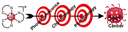
Liang He, Caiping Tan, Qian Cao, Zongwan Mao
2018, 30 (10): 1548-1556 | DOI: 10.7536/PC180610
Published: 15 October 2018
Abstract
Due to the excellent phosphorescent properties, the coordinatively saturated and substitutionally inert cyclometalated iridium(Ⅲ) complexes have been widely used in biological imaging and biosensing. In recent years, the application of this kind of iridium(Ⅲ) complexes in cancer treatment has attracted wide attention due to its anticancer efficacy and novel mechanisms. Recent advances of phosphorescent cyclometalated iridium(Ⅲ) complexes of the formula [Ir(C^N)2(N^N)]+ in anticancer chemotherapy and photodynamic therapy (PDT) are reviewed in this article. The iridium(Ⅲ) complexes targeting different organelles or as protein-protein interactions inhibitors or being applied in one-photon and two-photon PDT are summarized and discussed, which provides guidance for the development of new metal-based anticancer drugs. Finally, the development and application of phosphorescent cyclometalated iridium(Ⅲ) complexes in cancer treatment is discussed and prospected.
Contents
1 Introduction
2 Organelle-targeted phosphorescent anticancer cyclometalated iridium (Ⅲ) complexes
2.1 Mitochondria-targeted
2.2 Lysosome-targeted
2.3 Other organelles-targeted
3 Cyclometalated iridium(Ⅲ) complexes as protein-protein interactions inhibitors
4 Cyclometalated iridium(Ⅲ) complexes used in photodynamic therapy(PDT)
4.1 Mechanisms
4.2 One-photon PDT
4.3 Two-photon PDT
5 Conclusion and outlook
Multifunctional Nanodrug Delivery Systems for Platinum-Based Anticancer Drugs
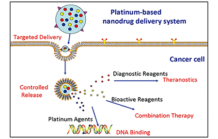
Juan Shen, Yang Zhu, Hongdong Shi, Yangzhong Liu
2018, 30 (10): 1557-1572 | DOI: 10.7536/PC180612
Published: 15 October 2018
Abstract
Platinum-based chemotherapy, especially cisplatin, is the standard first-line treatment for various types of cancer. However, the clinical application and development of platinum drugs have been greatly hampered by their severe adverse effects and inescapable drug resistance. The use of nanodrug delivery technology can effectively achieve targeted and controlled drug delivery and release. Moreover, nanodrug delivery technology can increase the bioavailability of platinum agents, reduce system toxicity and overcome the drug resistance. Therefore, it shows great potential for the treatment of cancer. In addition, the versatile nanocarriers facilitate the co-delivery of multiple agents with different bioactive functions, thereby providing possibilities for the combination therapy or theranostics in a single platform. Hence, nanodrug delivery systems present broad prospects for the precise cancer treatment. This article reviews the recent progress in the applications of multifunctional platinum-based nanodrug delivery systems for cancer therapy, and it consists of four aspects:targeted drug delivery, controlled drug release, combination therapy and theranostics. Meanwhile, the applications of new materials, new technologies and novel design ideas in platinum-based nanodrug delivery systems are also presented.
Contents
1 Introduction
2 Targeted drug delivery
2.1 Passive targeting based on EPR effect
2.2 Active targeting
3 Controlled drug release
3.1 pH-sensitive systems
3.2 Reduction-sensitive systems
3.3 Thermo-sensitive systems
3.4 Enzyme-sensitive systems
4 Combination therapy
4.1 Co-delivery of chemotherapeutic agents
4.2 Co-delivery of gene drugs and platinum agents
4.3 Co-delivery of phototherapy reagents and platinum agents
5 Theranostics
5.1 MRI-based theranostics
5.2 Optical imaging-based theranostics
5.3 Multimodal bioimaging-based theranostics
6 Conclusion and outlook
Application Prospect of Metal Complexes in Chemoimmunotherapy of Tumors
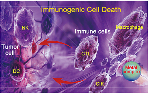
Yuewen Sun, Suxing Jin, Xiaoyong Wang, Zijian Guo
2018, 30 (10): 1573-1583 | DOI: 10.7536/PC180742
Published: 15 October 2018
Abstract
Tumor chemoimmunotherapy is a new method for the treatment of tumors through the combination of immunotherapy and chemotherapy taking advantage of synergistic effect. Metal drugs, represented by platinum agents, are important chemotherapeutic antitumor drugs with cross-linking DNA and preventing DNA replication as the mechanism of action. Nevertheless, these drugs have severe general toxicity and drug resistance. In recent years it was found that in addition to producing cytotoxicity, some metal complexes are involved in the immunomodulation by various mechanisms, including the most common induction of immunogenic cell death (ICD). This review introduces the basic concepts of tumor chemoimmunotherapy and the tumor microenvironment related to immunosuppression, outlines the immune activities and basic principles for immunomodulation of metal complexes like those of platinum. Finally, some non-platinum metal complexes with ICD-inducing potentials and other immunomodulating functions are described, and the existing problems and application potential of chemoimmunotherapy in the future are indicated. The combination of chemotherapy and immunotherapy not only makes use of the human immune system to enhance the antitumor effect of metal complexes, but also reduces the dose and toxic side effects of drugs, and therefore is one of the new directions for the design of metal-based antitumor drugs.
Contents
1 Introduction
1.1 Tumor immunotherapy and chemotherapy
1.2 Immunosuppressive tumor microenvironment
2 Metal complexes and chemoimmunotherapy
2.1 Immunocompetence of metal complexes
2.2 Immunomodulation effect of metal complexes
2.3 Immunogenic cell death
3 Chemoimmunotherapeutic effect of platinum-based drugs
3.1 Potential pathways for platinum-based drugs participating in immunomodulation
3.2 Platinum complexes with ICD-inducing effect
3.3 Chemoimmunotherapeutic platinum complexes
3.4 Combination of platinum-based drugs with other drugs for chemoimmunotherapy
4 Immunostimulation potential of non-platinum complexes
5 Conclusion and outlook
Inorganic Nanomaterials for Tumor Comprehensive Therapy
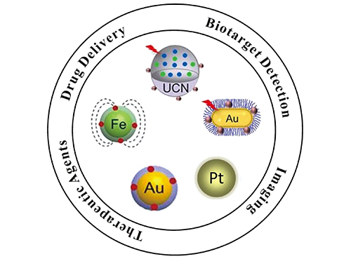
Jun Hu, Yuzhu Yao, Yanxiao Ao, Hai Yang, Xiangliang Yang*, Huibi Xu
2018, 30 (10): 1584-1591 | DOI: 10.7536/PC180616
Published: 15 October 2018
Abstract
Inorganic nanomaterials are widely applied in the diagnosis and comprehensive therapy of multiple diseases, in particular tumors, because of their unique nano-characters. Superparamagnetic iron oxide nanoparticles, upconversion nanoparticles and noble metal nanoparticles are the representative ones, possessing excellent magnetic, optical and thermal properties respectively. In this review, we focus on the applications of the three kinds of inorganic nanomaterials in bio-detection in vitro, molecular imaging in vivo, targeting anti-tumor drug delivery and tumor therapy. The advantages as well as the limitations of them in tumor theranostics are discussed. By this way, some references and suggestions are provided to develop more biocompatible inorganic nanomaterials with better theranostic effects, which will promote their clinical transformation.
Contents
1 Introduction
2 Superparamagnetic iron oxide nanoparticles
2.1 Separation and detection in vitro
2.2 Magnetic resonance imaging
2.3 Magnetic hyperthermia therapy
3 Upconversion nanoparticles
3.1 Multimodality imaging
3.2 Comprehensive therapy of tumor
4 Noble metal (Au、Pt) nanoparticles
5 Conclusion and outlook
Neurotoxicity of Key Metals in Parkinson's Disease
Hui Huang, Jun Chen, Huiru Lu, Mengxue Zhou, Yi Hu, Zhifang Chai
2018, 30 (10): 1592-1600 | DOI: 10.7536/PC180609
Published: 15 October 2018
Abstract
As China's aging population continues to increase, it is estimated that people with Parkinson's disease (PD) in China account for about half of PD cases in the world. Therefore, PD has become one of the fastest growing threats to public health in China. Currently, there is no cure for PD. The etiology and pathogenesis of PD remain elusive. Metal dyshomeostasis and oxidative stress are deemed as important risk factors for PD. Herein, recent advances in the neurotoxicity of key metals in PD are reviewed, and possible mechanisms underlying iron/copper-mediated neuron lesion are discussed. In addition, the application of synchrotron radiation in elemental analysis and mechanistic study of PD are briefly introduced. Lastly, the challenges and perspectives for bioinorganic chemistry in PD are discussed.
Contents
1 Introduction
2 The issues of bioinorganic chemistry in Parkinson's disease
2.1 Oxidative stress
2.2 Metal homeostasis
3 Iron in Parkinson's disease
3.1 Ferroptosis
3.2 Interaction between iron cations and proteins
3.3 Iron chelators
4 Copper in Parkinson's disease
4.1 Copper homeostasis
4.2 Interaction between cupric ions and α-synuclein
5 The application of synchrotron radiation-based elemental analysis to Parkinson's disease
6 Conclusion and outlook