中文
Announcement
More
Progress in Chemistry 2023, Vol. 35 Issue (5): 655-682 DOI: 10.7536/PC221024   Next Articles

• Review •

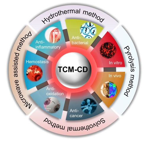
Synthesis of Traditional Chinese Medicines-Derived Carbon Dots for Bioimaging and Therapeutics

Jing He1,2, Jia Chen1,2(), Hongdeng Qiu1,2()   

  1. 1 Lanzhou Institute of Chemical Physics, Chinese Academy of Sciences,Lanzhou 730000, China
    2 University of Chinese Academy of Sciences,Beijing 101408, China
  • Received: Revised: Online: Published:
  • Contact: * e-mail: hdqiu@licp.cas.cn(Hongdeng Qiu);jiachen@licp.cas.cn(Jia Chen)
  • Supported by:
    CAS “Light of West China” Program(xbzg-zdsys-202008); Youth Innovation Promotion Association CAS(2021420)
Richhtml ( 106 ) PDF ( 1418 ) Cited
Export

EndNote

Ris

BibTeX

Carbon dots (CDs), with particle size less than 10 nm, are a new type of zero-dimensional photoluminescence nanomaterials. Due to the obvious advantages of adjustable fluorescence emission and excitation wavelength, light stability, low toxicity, good water solubility and biocompatibility, etc., CDs have been widely researched in recent years. As a treasure of ancient Chinese science, Traditional Chinese medicine (TCM) is rich in various active ingredients and plays a variety of pharmacodynamic effects, which has been used for thousands of years. TCM-CDs prepared with TCM as carbon source can create some special functions, and then may play a greater medicinal value. In this paper, the synthesis of TCM-CDs and its application in biological imaging and medical therapy are reviewed. Firstly, different synthetic methods of TCM-CDs (including hydrothermal, pyrolysis, solvothermal and microwave assisted method) are introduced in detail, and their advantages and disadvantages are compared. Subsequently, the latest research on TCM-CDs in biological imaging and medical treatment is comprehensively analyzed. This paper focuses on the application of imaging different types of cells in vitro and the distribution and uptake of TCM-CDs guided by imaging in vivo (mice, zebrafish, etc.). In addition, the intrinsic pharmacological activities of these TCM-CDs (including antibacterial, anti-inflammatory, hemostatic, antioxidant and anticancer, etc.) and their mechanisms are also discussed in order to improve and promote their clinical application. Finally, the importance of TCM-CDs research, the main problems and challenges in this fields and the future development direction are summarized and outlooked.

Contents

1 Introduction

2 Synthetic method of TCM-CDs

2.1 Hydrothermal method

2.2 Pyrolysis method

2.3 Solvothermal method

2.4 Microwave assisted method

3 Application of TCM-CDs in bioimaging

3.1 In vitro imaging

3.2 In vivo imaging

4 Application of TCM-CDs in therapeutics

4.1 Anti-bacterial

4.2 Anti-inflammatory

4.3 Hemostasis

4.4 Anti-oxidation

4.5 Anti-cancer

4.6 Other therapeutic effects

5 Conclusion and outlook

Fig.1 Schematic illustration of classifications and corresponding structures of CDs[2]
Fig. 2 Schematic illustration of synthesis route for N-CDs and the application in Ag+ sensing and RGB color analysis[28]
Fig. 3 (A) Schematic illustration for the synthesis and applications of fluorescent N-CDs[50]; (B) Schematic of the preparation and detection of Cr(Ⅵ) using CDs fluorescent probe[51]
Fig. 4 (A)Schematic illustration of the synthesis of fluorescent C-dots from peanut shells and their application in multicolor living cell imaging[54]; (B)Schematic synthesis of CDs from pyrolysis of plant leaves and the PL enhancement by plasma and microwave irradiation[55]; (C)Schematic representation of synthesis and applications of Durian Peel-based CDs[56]
Fig.5 Schematic of the preparation procedure and application of CP-CDs[63]
Fig. 6 Schematic illustration for the preparation of SC-CDs derived from alive silkworm chrysalis and the presentation of blue photoluminescence[67]
Table 1 Comparison of size, fluorescence properties and fluorescence QY of CDs prepared from different TCM sources, synthesis methods and conditions
Source Synthesis method Reaction temperature ( ℃/W) Reaction time (h) Exciation (nm) Emission (nm) Average particle size (nm) Quantum yield (%) Surface modification ref
Reynoutria japonica Houtt. Hydrothermal 200 3 320 400 35 11.5 - 24
Citrus Bergamot Hydrothermal 200 5 330 440 10 50.78 - 25
Perilla Frutescens(L.)Britt Hydrothermal 260 5 360 450 2.8 9.01 - 26
Lycium chinense Miller Hydrothermal 180 24 427 550 4.5 21.8 - 28
Lycium chinense Miller Hydrothermal 200 5 350 430 3.3 17.2 NH3·H2O 29
Zingiber officinale Roscoe Hydrothermal 300 2 325 400 4.3 13.4 - 38
Trapa bispinosa Roxb. Hydrothermal 90 2 450 520 7.5 1.2 - 39
Allium sativum L. Hydrothermal 200 3 360 442 10.7 17.5 - 40
Salvia miltiorrhiza Bunge Hydrothermal 100~180 6 400 490 1.53~16.94 30~40 - 44
Mentha haplocalyx Briq. Hydrothermal 200 5 360 450 7 7.64 - 46
Mentha haplocalyx Briq. Hydrothermal 180 8 363 441 5 4.5 - 47
Aloe vera Hydrothermal 180 11 441 503 5 10.3 - 48
Brassica oleracea Linnaeus var. capitata Linnaeus Hydrothermal 140 5 345 432 4 12.5 - 49
Lilium brownii F. E. Brown var. Colchesteri Wils. Hydrothermal 240 12 340 405 4 11 - 50
Poria cocos (Schw.) Wolf Hydrothermal 200 5 376 450 4 4.8 - 51
Ginkgo biloba Linn. Hydrothermal 160~200 8 420 520 3.81 3.33 - 69
Salvia miltiorrhiza Bunge Hydrothermal 150 6 420 526 3.32 - - 74
Charred Triplet Hydrothermal 100 2 340 447 5.1 7.95 - 131
Litchi chinensis Sonn. Pyrolysis 300 2 365 440 1.12 10.6 - 53
Peanut Shells Pyrolysis 250 2 320 440 1.6 9.91 - 54
Durian Peel Pyrolysis 250 5 368 480 10 11 - 56
Gynostemma pentaphyllum
(Thunb.) Makino
Pyrolysis 400 4 320 400 2.49 5.7 - 57
Papaya Solvothermal 200 5 370 450 3.4/10.8 18.98/18.39 ethanol 60
Saccharum sinensis Roxb. Solvothermal 250 6 350 430 1 10.7 ethanol 61
Saccharum sinensis Roxb. Solvothermal 120 8 360 460 5 - Urea and ethanol 62
Codonopsis pilosula Solvothermal 25 4 390 456 11.54 12.8 methanol 63
Mentha canadensis Linnaeus Microwave 960 0.07~0.17 340 436 2.43 17 - 66
Bombyx mori L. Microwave 210 0.75 350 440 19 46 - 67
Zingiberis rhizome and
Alpinia officinarum
Microwave 450 0.08~0.67 - - 10 - - 68
Ginkgo biloba Linn. Microwave 800 0.08~0.25 440 550 2.82 0.65 - 69
Panax ginseng Microwave 700 0.5 380 500 2 8 AgNPs 70
Talinum paniculatum (Jacq.)
Gaertn.
Microwave 700 0.5 380 470 2 - Rutin 107
Fig. 7 Confocal microscope images of A549 cells treated with FCP-B, FCP-G and FCP-Y, observed during a 4 h incubation period[79]
Fig. 8 High-content imaging analysis of cell apoptosis and necrosis by hoechst/PI double-staining method[89]
Fig. 9 Bioimaging of bacteria in presence of CDs in blue, yellow and red region (A) E. Coli and (B) B.Subtilis[92]
Fig. 10 Imaging and sensing of intracellular CR based on Mis-mPD-CDs. CLSM images of A549, C. albicans, E. coli, and S. aureus, and treated with CR of different concentrations and then incubated with Mis-mPD-CDs for 30 min[93]
Fig. 11 In vivo biodistribution of FCP-B, FCP-G and FCP-Y in balb/c nude mice after tail vein injection of 5 mg·kg-1 of body weight. The in vivo biodistribution and corresponding intensities of FCP-B (a, b), FCP-G (c, d) and FCP-Y (e, f) respectively
Fig. 12 One-photon and two-photon bioimaging of CDs. (A) In vivo imaging of supine nude mice with intravenous injection of CDs at different time points (Ⅰ: thoracic region, Ⅱ: area of liver, Ⅲ: area of small intestine, Ⅳ: area of large intestine, Ⅴ: bladder region). (B)Real-time ex vivo imaging of nude mice with intravenous injection of CDs at different time points
Fig. 13 Real-time NIR fluorescence images of the mice tumor with NB-CDs at different time points[88]
Fig. 14 In vivo imaging of male BALB/c mice after injection of Pn N-CDs at different time intervals[98]
Fig. 15 The photoluminescence decay of CDs in zebrafish. The fluorescent microscopic images of bright field and fluorescent field of zebrafish embryos after exposure to 0.4 mg/mL HCDs and SCDs solutions for 2 days at different time points[100]
Table 2 The application of TCM-CDs in bioimaging
Source FL color Applied Ex/Em (nm) Application Biotarget ref
Citrus junos Tanada blue, green 405, 488/- Imaging of cells MG-63 74
Mentha haplocalyx Briq blue, green, red 360, 470, 530/447,
525, 593
Imaging of cells MCF-7 75
Prunus persica blue - Imaging of cells MDA-MB-231 76
Prunus mume blue, green 365/- Imaging of cells MDA-MB-231 77
Curcuma Longa blue, yellow, red 405, 488, 543/- Imaging of cells KB 78
Mangifera indica L. blue, green, yellow 488, 488, 513/505,
530, 560
Imaging of cells A549 79
Allium sativum L. blue, green, yellow 385, 480,550/- Imaging of cells A549 40
Coriandrum sativum Linn. green 470/525 Imaging of cells L-132 80
Dendranthema morifolium blue, green 405, 488/- Imaging of cells HeLa 81
Mentha haplocalyx Briq. blue, yellow, red 380, 480, 590/- Imaging of cells HeLa 82
Brassica compestris L.var.purpurea Bailey blue, yellow, red 405, 488, 559/- Imaging of cells HeLa 83
Bombyx mori L. blue, yellow, red 340, 495, 550/- Imaging of cells HeLa 67
Abelmoschus esculentus (Linn.) Moench blue 340/410 Imaging of cells HeLa 84
Lycium chinense Miller blue, glaucous, green 400, 415, 485/- Imaging of cells HeLa 29
Alisma plantago-aquatica Linn. blue, yellow, red 340, 460, 520/- Imaging of cells HeLa 85
Ginkgo biloba Linn. blue, green 405, 488/- Imaging of cells HeLa, KYSE-410 69
Benincasa hispida (Thunb.) Cogn. blue 365/- Imaging of cells HepG2 86
Prunus cerasifera Ehrhart f. atropurpurea (Jacq.) Rehd. blue, green 405, 488/- Imaging of cells HepG2 87
Litchi chinensis Sonn blue 405/- Imaging of cells HepG2 88
Ginsenoside Re blue to red 360-530/- Imaging of cells A375 89
Brassica oleracea Linnaeus var.
capitata Linnaeus
blue, green, red 405, 488, 543/- Imaging of cells HaCaT 49
Phyllanthus acidus (L.) Skeels blue, green, red 405, 488, 555/- Imaging of cells Clone9 90
Cymbopogon citratus (D. C.) Stapf blue, yellow, red - Imaging of bacteria BY4742 91
Ocimum sanctum Linn. blue, yellow, red 405, 488, 561/- Imaging of bacteria B. subtilis,
E. coli
92
Salvadora persica green 488/550 Imaging of bacteria C. albicans,E. coli, S. aureus 93
Allium cepa Linn. green - Imaging of bacteria E.coli, S.aureus 94
Mangifera indica L. blue to yellow - In vivo imaging mice 79
Taxus chinensis (Pilger) Rehd. red 640/705 In vivo imaging of tumor mice 97
Litchi chinensis Sonn. blue to red - In vivo imaging of tumor mice 88
Panax notoginseng blue to red - In vivo imaging of tumor BALB/c mice 98
Pisum sativum Linn. blue - In vivo imaging mice 99
Crinis Carbonisatus blue, green, red 385, 480, 550/- In vivo imaging Zebrafish 100
Gynostemma pentaphyllum
(Thunb.) Makino
blue, green, red - In vivo imaging Zebrafish 57
Salvadora persica green 488/550 In vivo imaging Zebrafish 93
Panax notoginseng blue, yellow, red 405, 488, 543/- In vivo imaging P. caudatum 98
Fig. 16 General bactericidal mechanisms of action of CDs. (A) Schematic representation of the initial electrostatic interaction between CDs and the bacterial cell wall. (B) CDs internalization, intercalation in the bacterial membrane, and irreversible disruption with a leak of cytoplasmatic material. (C) CD-promoted bacterial photodynamic inactivation with ROS production and DNA damage[105]
Fig. 17 SEM images of E. coli: (A~C) light irradiation without MCDs, (D~F) MCDs treated under dark conditions, and (G~I) MCDs treated with visible light irradiation for 12 h[106]
Fig. 18 (a) Schematic illustration of the effect of the onion carbon dots on the integrity of the bacterial cell walls and membranes. Effect of onion carbon dots on the release of extracellular (b) AKP and (c) ATP content in Pseudomonas fragi[94]
Fig. 19 Schematic illustration of CDs for ROS depletion and anti-inflammation therapy[108]
Fig. 20 Macroscopic images of ASAC-CDs ameliorating an LPS-induced acute lung injury (ALI) in rats. (A) Normal saline group; (B) model group; (C) positive control group; (D) high-dose ASAC-CDs group; (E) medium-dose ASAC-CDs group; and (F) low-dose ASAC-CDs group[112]
Fig. 21 Schematic of the Mechanism of CDs Alleviating the Oxidative Damage of Italian Lettuce under Salt Stress[44]
Fig. 22 Schematic of the function of J-CDs[132]
[1]
Xu X Y, Ray R, Gu Y L, Ploehn H J, Gearheart L, Raker K, Scrivens W A. J. Am. Chem. Soc., 2004, 126(40): 12736.

doi: 10.1021/ja040082h
[2]
Ai L, Shi R, Yang J, Zhang K, Zhang T R, Lu S Y. Small, 2021, 17(48): 2007523.

doi: 10.1002/smll.v17.48
[3]
Fatahi Z, Esfandiari N, Ranjbar Z. J. Anal. Test., 2020, 4(4): 307.

doi: 10.1007/s41664-020-00149-6
[4]
Tang Z J, Li G K, Hu Y L. Prog. Chem., 2016, 28: 1455.
(唐志姣, 李攻科, 胡玉玲. 化学进展, 2016, 28: 1455.).

doi: 10.7536/PC160504
[5]
Li C H, Liu Y M, Lu B, Sana U, Ren X Y, Sun Y P. Prog. Chem., 2022, 34(3): 499.
(李程浩, 刘亚敏, 卢彬, 萨拉乌拉, 任先艳, 孙亚平. 化学进展, 2022, 34(3): 499.).

doi: 10.7536/PC210303
[6]
Zhai Y P, Zhang B W, Shi R, Zhang S Y, Liu Y, Wang B Y, Zhang K, Waterhouse G I N, Zhang T R, Lu S Y. Adv. Energy Mater., 2022, 12(6): 2103426.

doi: 10.1002/aenm.v12.6
[7]
Semeniuk M, Yi Z H, Poursorkhabi V, Tjong J, Jaffer S, Lu Z H, Sain M. ACS Nano, 2019, 13(6): 6224.

doi: 10.1021/acsnano.9b00688 pmid: 31145587
[8]
Wei Y Y, Chen L, Wang J L, Yu S P, Liu X G, Yang Y Z. Prog. Chem., 2020, 32: 381.
(卫迎迎, 陈琳, 王军丽, 于世平, 刘旭光, 杨永珍. 化学进展, 2020, 32: 381.).

doi: 10.7536/PC190739
[9]
Hutton G A M, Martindale B C M, Reisner E. Chem. Soc. Rev., 2017, 46(20): 6111.

doi: 10.1039/C7CS00235A
[10]
Xiong W, Zhao Y, Cheng J J, Zhang M L, Sun Z W, Luo J, Zhu Y F, Zhang Y X, Kong H, Qu H H. Chinese Traditional and Herbal Drugs, 2019, 50: 1388.
(熊威, 赵琰, 成金俊, 张美龄, 孙紫薇, 罗娟, 朱雅凡, 张亚雪, 孔慧, 屈会化. 中草药, 2019, 50: 1388.).
[11]
He C, E S, Yan H H, Li X J. Prog. Chem., 2022, 34: 356.
(何闯, 鄂爽, 闫鸿浩, 李晓杰. 化学进展, 2022, 34: 356.).

doi: 10.7536/PC210105
[12]
Chen S T, Chen C Q, Wang J, Luo F, Guo L H, Qiu B, Lin Z Y. J. Anal. Test., 2021, 5(1): 84.

doi: 10.1007/s41664-021-00162-3
[13]
Wang J L, Wang Y L, Zheng J X, Yu S P, Yang Y Z, Liu X G. Prog. Chem., 2018, 30: 1186.
(王军丽, 王亚玲, 郑静霞, 于世平, 杨永珍, 刘旭光. 化学进展, 2018, 30: 1186.).

doi: 10.7536/PC180103
[14]
Sharma A, Das J. J. Nanobiotechnology, 2019, 17(1): 92.

doi: 10.1186/s12951-019-0525-8
[15]
Zhang H J, Qiao X, Cai T P, Chen J, Li Z, Qiu H D. Anal. Bioanal. Chem., 2017, 409(9): 2401.

doi: 10.1007/s00216-017-0187-z
[16]
Yang Y L, Zhang H J, Chen J, Li Z, Zhao L, Qiu H D. Analyst, 2020, 145(3): 1056.

doi: 10.1039/C9AN02246E
[17]
Chen J, Gong Z J, Tang W Y, Row K H, Qiu H D. Trac Trends Anal. Chem., 2021, 134: 116135.

doi: 10.1016/j.trac.2020.116135
[18]
Chen J, Yuan N, Jiang D N, Lei Q, Liu B, Tang W Y, Row K H, Qiu H D. Chin. Chem. Lett., 2021, 32(11): 3398.

doi: 10.1016/j.cclet.2021.04.052
[19]
Egorova M N, Tomskaya A E, Kapitonov A N, Alekseev A A, Smagulova S A. J. Struct. Chem., 2018, 59(4): 780.

doi: 10.1134/S0022476618040054
[20]
Liu Y S, Zhao Y N, Zhang Y Y. Sens. Actuat. B Chem., 2014, 196: 647.

doi: 10.1016/j.snb.2014.02.053
[21]
Kong L Y, Tan R X. Nat. Prod. Rep., 2015, 32(12): 1617.

doi: 10.1039/C5NP00133A
[22]
Miao R, Meng Q, Wang C, Yuan W. Evid. Based Complementary Altern. Med., 2022, 2022: 8075344.
[23]
Zhang Y, Wang S N, Lu F, Zhang M L, Kong H, Cheng J J, Luo J, Zhao Y, Qu H H. J. Nanobiotechnology, 2021, 19(1): 257.

doi: 10.1186/s12951-021-00908-2
[24]
Wu D, Huang X M, Deng X, Wang K, Liu Q Y. Anal. Methods, 2013, 5(12): 3023.

doi: 10.1039/c3ay40337h
[25]
Yu J, Song N, Zhang Y K, Zhong S X, Wang A J, Chen J R. Sens. Actuat. B Chem., 2015, 214: 29.

doi: 10.1016/j.snb.2015.03.006
[26]
Zhao X Y, Liao S, Wang L M, Liu Q, Chen X Q. Talanta, 2019, 201: 1.

doi: 10.1016/j.talanta.2019.03.095
[27]
Zuo G C, Xie A M, Li J J, Su T, Pan X H, Dong W. J. Phys. Chem. C, 2017, 121(47): 26558.

doi: 10.1021/acs.jpcc.7b10179
[28]
Tang S Y, Chen D, Guo G Q, Li X M, Wang C X, Li T T, Wang G. Sci. Total Environ., 2022, 825: 153913.

doi: 10.1016/j.scitotenv.2022.153913
[29]
Sun X H, He J, Yang S H, Zheng M D, Wang Y Y, Ma S, Zheng H P. J. Photochem. Photobiol. B Biol., 2017, 175: 219.

doi: 10.1016/j.jphotobiol.2017.08.035
[30]
Sharma V, Tiwari P, Mobin S M. J. Mater. Chem. B, 2017, 5(45): 8904.

doi: 10.1039/c7tb02484c pmid: 32264117
[31]
Humaera N A, Fahri A N, Armynah B, Tahir D. Luminescence, 2021, 36(6): 1354.

doi: 10.1002/bio.v36.6
[32]
Luo W K, Zhang L L, Yang Z Y, Guo X H, Wu Y, Zhang W, Luo J K, Tang T, Wang Y. J. Nanobiotechnology, 2021, 19(1): 320.

doi: 10.1186/s12951-021-01072-3
[33]
Li D, Xu K Y, Zhao W P, Liu M F, Feng R, Li D Q, Bai J, Du W L. Front. Pharmacol., 2022, 13: 815479.

doi: 10.3389/fphar.2022.815479
[34]
Wu X C, Liang W H, Cai C X. Prog. Chem., 2021, 33: 1059.
(吴星辰, 梁文慧, 蔡称心. 化学进展, 2021, 33: 1059.).

doi: 10.7536/PC200715
[35]
Yan F Y, Zhou Y, Wang M, Dai L F, Zhou X G, Chen L. Prog. Chem., 2014, 26: 61.
(颜范勇, 邹宇, 王猛, 代林枫, 周旭光, 陈莉. 化学进展, 2014, 26: 61.).

doi: 10.7536/PC130643
[36]
Liu Y S, Li W, Wu P, Liu S X. Prog. Chem., 2018, 30: 349.
(刘禹杉, 李伟, 吴鹏, 刘守新. 化学进展, 2018, 30: 349.).

doi: 10.7536/PC170808
[37]
Rong M C, Wang D R, Li Y Y, Zhang Y Z, Huang H Y, Liu R F, Deng X Z. J. Anal. Test., 2021, 5(1): 51.

doi: 10.1007/s41664-021-00161-4
[38]
Li C L, Ou C M, Huang C C, Wu W C, Chen Y P, Lin T E, Ho L C, Wang C W, Shih C C, Zhou H C, Lee Y C, Tzeng W F, Chiou T J, Chu S T, Cang J S, Chang H T. J. Mater. Chem. B, 2014, 2(28): 4564.

doi: 10.1039/c4tb00216d pmid: 32261557
[39]
Mewada A, Pandey S, Shinde S, Mishra N, Oza G, Thakur M, Sharon M, Sharon M,. Mater. Sci. Eng. C, 2013, 33(5): 2914.

doi: 10.1016/j.msec.2013.03.018
[40]
Zhao S J, Lan M H, Zhu X Y, Xue H T, Ng T W, Meng X M, Lee C S, Wang P F, Zhang W J. ACS Appl. Mater. Interfaces, 2015, 7(31): 17054.

doi: 10.1021/acsami.5b03228
[41]
Zhu S J, Meng Q N, Wang L, Zhang J H, Song Y B, Jin H, Zhang K, Sun H C, Wang H Y, Yang B. Angew. Chem. Int. Ed., 2013, 52(14): 3953.

doi: 10.1002/anie.v52.14
[42]
Bhattacharyya S, Ehrat F, Urban P, Teves R, Wyrwich R, Döblinger M, Feldmann J, Urban A S, Stolarczyk J K. Nat. Commun., 2017, 8: 1401.

doi: 10.1038/s41467-017-01463-x pmid: 29123091
[43]
Kwon W, Lee G, Do S, Joo T, Rhee S W. Small, 2014, 10(3): 506.

doi: 10.1002/smll.v10.3
[44]
Li Y J, Li W, Yang X, Kang Y Y, Zhang H R, Liu Y L, Lei B F. ACS Appl. Nano Mater., 2021, 4(1): 113.

doi: 10.1021/acsanm.0c02419
[45]
Yuan F L, Wang Z B, Li X H, Li Y C, Tan Z A, Fan L Z, Yang S H. Adv. Mater., 2017, 29(3): 1604436.

doi: 10.1002/adma.v29.3
[46]
Shen Y L, Wu H Y, Wu W H, Zhou L, Dai Z F, Dong S P. Mater. Express, 2020, 10(7): 1135.

doi: 10.1166/mex.2020.1745
[47]
Raveendran V, Suresh Babu A R, Renuka N K. RSC Adv., 2019, 9(21): 12070.

doi: 10.1039/c9ra02120e
[48]
Xu H, Yang X P, Li G, Zhao C, Liao X J. J. Agric. Food Chem., 2015, 63(30): 6707.

doi: 10.1021/acs.jafc.5b02319
[49]
Alam A M, Park B Y, Ghouri Z K, Park M, Kim H Y. Green Chem., 2015, 17(7): 3791.

doi: 10.1039/C5GC00686D
[50]
Atchudan R, Edison T N J I, Aseer K R, Perumal S, Lee Y R. Colloids Surf. B Biointerfaces, 2018, 169: 321.

doi: 10.1016/j.colsurfb.2018.05.032
[51]
Huang Q Q, Bao Q Q, Wu C Y, Hu M R, Chen Y N, Wang L, Chen W D. J. Pharm. Anal., 2022, 12(1): 104.

doi: 10.1016/j.jpha.2021.04.004
[52]
Wang S, Wu S H, Fang W L, Guo X F, Wang H. Dyes Pigments, 2019, 164: 7.

doi: 10.1016/j.dyepig.2019.01.004
[53]
Xue M Y, Zou M B, Zhao J J, Zhan Z H, Zhao S L. J. Mater. Chem. B, 2015, 3(33): 6783.

doi: 10.1039/C5TB01073J
[54]
Xue M Y, Zhan Z H, Zou M B, Zhang L L, Zhao S L. New J. Chem., 2016, 40(2): 1698.

doi: 10.1039/C5NJ02181B
[55]
Zhu L L, Yin Y J, Wang C F, Chen S. J. Mater. Chem. C, 2013, 1(32): 4925.

doi: 10.1039/c3tc30701h
[56]
Praneerad J, Neungnoraj K, In I, Paoprasert P. Key Eng. Mater., 2019, 803: 115.

doi: 10.4028/www.scientific.net/KEM.803
[57]
Wei X J, Li L, Liu J L, Yu L D, Li H B, Cheng F, Yi X T, He J M, Li B S. ACS Appl. Mater. Interfaces, 2019, 11(10): 9832.

doi: 10.1021/acsami.9b00074
[58]
Aji M P, Susanto, Wiguna P A, Sulhadi. J. Theor. Appl. Phys., 2017, 11(2): 119.

doi: 10.1007/s40094-017-0250-3
[59]
Chen Y Q, Lian H Z, Wei Y, He X, Chen Y, Wang B, Zeng Q G, Lin J. Nanoscale, 2018, 10(14): 6734.

doi: 10.1039/C8NR00204E
[60]
Wang N, Wang Y, Guo T, Yang T, Chen M, Wang J. Biosens. Bioelectron., 2016, 85: 68.

doi: 10.1016/j.bios.2016.04.089
[61]
Moreira V A, Toito Suarez W, de Oliveira Krambeck Franco M, Gambarra Neto F F. Anal. Methods, 2018, 10(37): 4570.

doi: 10.1039/C8AY00928G
[62]
Sim L C, Wong J L, Hak C H, Tai J Y, Leong K H, Saravanan P. Beilstein J. Nanotechnol., 2018, 9: 353.

doi: 10.3762/bjnano.9.35
[63]
Qiu Y, Gao D, Yin H G, Zhang K L, Zeng J, Wang L J, Xia L, Zhou K, Xia Z N, Fu Q F. Sens. Actuat. B Chem., 2020, 324: 128722.

doi: 10.1016/j.snb.2020.128722
[64]
Shi Y P, Liu J J, Zhang Y, Bao J F, Cheng J L, Yi C Q. Chin. Chem. Lett., 2021, 32(10): 3189.

doi: 10.1016/j.cclet.2021.03.022
[65]
Chatzimitakos T, Kasouni A, Sygellou L, Leonardos I, Troganis A, Stalikas C. Sens. Actuat. B Chem., 2018, 267: 494.

doi: 10.1016/j.snb.2018.04.059
[66]
Architha N, Ragupathi M, Shobana C, Selvankumar T, Kumar P, Lee Y S, Kalai Selvan R. Environ. Res., 2021, 199: 111263.

doi: 10.1016/j.envres.2021.111263
[67]
Feng J, Wang W J, Hai X, Yu Y L, Wang J H. J. Mater. Chem. B, 2016, 4(3): 387.

doi: 10.1039/c5tb01999k pmid: 32263205
[68]
Isnaeni, Rahmawati I, Intan R, Zakaria M. J. Phys.: Conf. Ser., 2018, 985: 012004.
[69]
Li L L, Li L Y, Chen C P, Cui F L. Inorg. Chem. Commun., 2017, 86: 227.

doi: 10.1016/j.inoche.2017.10.006
[70]
Tejwan N, Sharma A, Thakur S, Das J. Inorg. Chem. Commun., 2021, 134: 108985.

doi: 10.1016/j.inoche.2021.108985
[71]
de Medeiros T V, Manioudakis J, Noun F, Macairan J R, Victoria F, Naccache R. J. Mater. Chem. C, 2019, 7(24): 7175.

doi: 10.1039/c9tc01640f
[72]
Singh R K, Kumar R, Singh D P, Savu R, Moshkalev S A. Mater. Today Chem., 2019, 12: 282.

doi: 10.1016/j.mtchem.2019.03.001
[73]
Lu M C, Duan Y X, Song Y H, Tan J S, Zhou L. J. Mol. Liq., 2018, 269: 766.

doi: 10.1016/j.molliq.2018.08.101
[74]
Sahu S, Behera B, Maiti T K, Mohapatra S. Chem. Commun., 2012, 48(70): 8835.

doi: 10.1039/c2cc33796g
[75]
Shahid S, Mohiyuddin S, Packirisamy G. J. Nanosci. Nanotechnol., 2020, 20(10): 6305.

doi: 10.1166/jnn.2020.17899 pmid: 32384980
[76]
Atchudan R, Edison T N J I, Lee Y R. J. Colloid Interface Sci., 2016, 482: 8.

doi: 10.1016/j.jcis.2016.07.058
[77]
Atchudan R, Edison T N J I, Sethuraman M G, Lee Y R. Appl. Surf. Sci., 2016, 384: 432.

doi: 10.1016/j.apsusc.2016.05.054
[78]
Mazrad Z A I, Kang E B, In I, Park S Y. Luminescence, 2018, 33(1): 40.

doi: 10.1002/bio.3370
[79]
Jeong C J, Roy A K, Kim S H, Lee J E, Jeong J H, In I, Park S Y. Nanoscale, 2014, 6(24): 15196.

doi: 10.1039/c4nr04805a pmid: 25375199
[80]
Sachdev A, Gopinath P. Anal., 2015, 140(12): 4260.

doi: 10.1039/C5AN00454C
[81]
Chai C F, Qiao X H, Zheng L L, Duan H B, Bian W, Choi M M F. Fuller. Nanotub. Carbon Nanostructures, 2022, 30(5): 534.

doi: 10.1080/1536383X.2021.1966419
[82]
Raveendran V, Kizhakayil R N. ACS Omega, 2021, 6(36): 23475.

doi: 10.1021/acsomega.1c03481 pmid: 34549145
[83]
Li L S, Jiao X Y, Zhang Y, Cheng C, Huang K, Xu L. Sens. Actuat. B Chem., 2018, 263: 426.

doi: 10.1016/j.snb.2018.02.141
[84]
Wan Y Y, Wang M, Zhang K L, Fu Q F, Gao M J, Wang L J, Xia Z N, Gao D. Microchem. J., 2019, 148: 385.

doi: 10.1016/j.microc.2019.05.026
[85]
Liu M J, Hao X L, Dai S J, Wang S Y, Wang Y, Zhang H. Chem. Res. Chin. Univ., 2022, 39: 234.

doi: 10.1007/s40242-022-2142-6
[86]
Feng X,. Jiang Y, Zhao J. RSC Adv., 2015, 5: 31250.

doi: 10.1039/C5RA02271A
[87]
Ma H P, Sun C C, Xue G, Wu G L, Zhang X H, Han X, Qi X H, Lv X, Sun H J, Zhang J B. Spectrochimica Acta A Mol. Biomol. Spectrosc., 2019, 213: 281.
[88]
Xue M Y, Zhao J J, Zhan Z H, Zhao S L, Lan C Q, Ye F G, Liang H. Nanoscale, 2018, 10(38): 18124.

doi: 10.1039/C8NR05017A
[89]
Yao H, Li J, Song Y B, Zhao H, Wei Z H, Li X Y, Jin Y R, Yang B, Jiang J L. Int. J. Nanomed., 2018, 13: 6249.

doi: 10.2147/IJN
[90]
Atchudan R, Edison T N J I, Aseer K R, Perumal S, Karthik N, Lee Y R. Biosens. Bioelectron., 2018, 99: 303.

doi: S0956-5663(17)30534-1 pmid: 28780346
[91]
Thota S P, Thota S M, Srimadh Bhagavatham S, Sai Manoj K, Sai Muthukumar V S, Venketesh S, Vadlani P V, Belliraj S K. IET Nanobiotechnol., 2018, 12(2): 127.

doi: 10.1049/nbt2.v12.2
[92]
Bhatt S, Bhatt M, Kumar A, Vyas G, Gajaria T, Paul P. Colloids Surf. B Biointerfaces, 2018, 167: 126.

doi: 10.1016/j.colsurfb.2018.04.008
[93]
Durrani S, Zhang J, Yang Z, Pang A P, Zeng J, Sayed S M, Khan A, Zhang Y Q, Wu F G, Lin F M. Anal. Chim. Acta, 2022, 1202: 339672.

doi: 10.1016/j.aca.2022.339672
[94]
Lin R, Cheng S S, Tan M Q. Food Funct., 2022, 13(4): 2098.

doi: 10.1039/D1FO03426J
[95]
Yang S T, Cao L, Luo P G, Lu F S, Wang X, Wang H F, Meziani M J, Liu Y F, Qi G, Sun Y P. J. Am. Chem. Soc., 2009, 131(32): 11308.

doi: 10.1021/ja904843x
[96]
Luo P G, Sahu S, Yang S T, Sonkar S K, Wang J P, Wang H F, LeCroy G E, Cao L, Sun Y P. J. Mater. Chem. B, 2013, 1(16): 2116.

doi: 10.1039/c3tb00018d
[97]
Liu J J, Geng Y J, Li D W, Yao H, Huo Z P, Li Y F, Zhang K, Zhu S J, Wei H T, Xu W Q, Jiang J L, Yang B. Adv. Mater., 2020, 32(17): 1906641.

doi: 10.1002/adma.v32.17
[98]
Zheng X D, Qin K H, He L P, Ding Y F, Luo Q, Zhang C T, Cui X M, Tan Y, Li L N, Wei Y L. Analyst, 2021, 146(3): 911.

doi: 10.1039/D0AN01599G
[99]
Su Q, Gan L L, Liu J, Yang X M. Anal. Biochem., 2020, 589: 113476.

doi: 10.1016/j.ab.2019.113476
[100]
Zhang J H, Niu A P, Li J, Fu J W, Xu Q, Pei D S. Sci. Rep., 2016, 6: 37860.

doi: 10.1038/srep37860
[101]
Phan L M T, Cho S. Bioinorg. Chem. Appl., 2022, 2022: 9303703.
[102]
Sun Y N, Zhang M, Bhandari B, Yang C H. Food Rev. Int., 2022, 38(7): 1513.

doi: 10.1080/87559129.2020.1818255
[103]
Dong X L, Liang W X, Meziani M J, Sun Y P, Yang L J. Theranostics, 2020, 10(2): 671.

doi: 10.7150/thno.39863
[104]
Wu Y Y, Li C, van der Mei H C, Busscher H J, Ren Y J. Antibiotics, 2021, 10(6): 623.

doi: 10.3390/antibiotics10060623
[105]
Ghirardello M, Ramos-Soriano J, Galan M C. Nanomaterials, 2021, 11(8): 1877.

doi: 10.3390/nano11081877
[106]
Venkateswarlu S, Viswanath B, Reddy A S, Yoon M. Sens. Actuat. B Chem., 2018, 258: 172.

doi: 10.1016/j.snb.2017.11.044
[107]
Tejwan N, Kundu M, Sharma A, Das J, Sil P C. Research Square,Preprint, (2020-05-19). DOI:10.21203/rs.3.rs-28922/v1.

doi: 10.21203/rs.3.rs-28922/v1
[108]
Kong B, Yang T, Cheng F, Qian Y, Li C M, Zhan L, Li Y F, Zou H Y, Huang C Z. J. Colloid Interface Sci., 2022, 611: 545.

doi: 10.1016/j.jcis.2021.12.107
[109]
Wang S N, Zhang Y, Kong H, Zhang M L, Cheng J J, Wang X K, Lu F, Qu H H, Zhao Y. Nanomedicine, 2019, 14(22): 2925.

doi: 10.2217/nnm-2019-0255
[110]
Wang X K, Zhang Y, Kong H, Cheng J J, Zhang M L, Sun Z W, Wang S N, Liu J X, Qu H H, Zhao Y. Artif. Cells Nanomed. Biotechnol., 2020, 48(1): 68.

doi: 10.1080/21691401.2019.1699810
[111]
Zhang M L, Cheng J J, Hu J, Luo J, Zhang Y, Lu F, Kong H, Qu H H, Zhao Y. J. Nanobiotechnology, 2021, 19(1): 105.

doi: 10.1186/s12951-021-00847-y
[112]
Zhao Y S, Zhang Y, Kong H, Cheng G L, Qu H H, Zhao Y. Int. J. Nanomed., 2022, 17: 1.

doi: 10.2147/IJN.S338886
[113]
Wu J S, Zhang M L, Cheng J J, Zhang Y, Luo J, Liu Y H, Kong H, Qu H H, Zhao Y. Int. J. Nanomed., 2020, 15: 4139.

doi: 10.2147/IJN.S248467
[114]
Zhao Y S, Li W Y, Cao T Y, Zhao Y, Qu H H, Guid. J.. Tradit. Chin. Med. Pharm, 2021, 27: 56.
(赵玉升, 李伟洋, 曹天佑, 赵琰, 屈会化. 中医药导报, 2021, 27: 56.).
[115]
Guo Y M, Zhang L F, Cao F P, Leng Y M. Sci. Rep., 2016, 6: 35795.

doi: 10.1038/srep35795
[116]
Zhang M, Zhao Y, Cheng J, Liu X, Wang Y, Yan X, Zhang Y, Lu F, Wang Q, Qu H. Artificial Cells, Nanomedicine, and Biotechnology, 2018, 46: 1562.
[117]
Wang Y Z, Kong H, Liu X M, Luo J, Xiong W, Zhu Y F, Zhang Y X, Zhang Y H, Zhao Y, Qu H H. J. Biomed. Nanotechnol., 2018, 14(9): 1635.

doi: 10.1166/jbn.2018.2613
[118]
Sun Z W, Lu F, Cheng J J, Zhang M L, Zhang Y, Xiong W, Zhao Y, Qu H H. Artif. Cells Nanomed. Biotechnol., 2018, 46(sup3): 308.

doi: 10.1080/21691401.2018.1492419
[119]
Yan X, Zhao Y, Luo J, Xiong W, Liu X M, Cheng J J, Wang Y Z, Zhang M L, Qu H H. J. Nanobiotechnology, 2017, 15(1): 60.

doi: 10.1186/s12951-017-0296-z
[120]
Luo J, Kong H, Zhang M L, Cheng J J, Sun Z W, Xiong W, Zhu Y F, Zhao Y, Qu H H. J. Biomed. Nanotechnol., 2019, 15(1): 151.

doi: 10.1166/jbn.2019.2675
[121]
Liu X M, Wang Y Z, Yan X, Zhang M L, Zhang Y, Cheng J J, Lu F, Qu H H, Wang Q G, Zhao Y. Nanomedicine, 2018, 13(4): 391.

doi: 10.2217/nnm-2017-0297
[122]
Luo J, Zhang M L, Cheng J J, Wu S H, Xiong W, Kong H, Zhao Y, Qu H H. RSC Adv., 2018, 8(66): 37707.

doi: 10.1039/C8RA06340K
[123]
Chen Z, Ye S Y, Yang Y, Li Z Y. Pharm. Biol., 2019, 57(1): 498.

doi: 10.1080/13880209.2019.1645700 pmid: 31401925
[124]
Cheng J J, Zhang M L, Sun Z W, Lu F, Xiong W, Luo J, Kong H, Wang Q G, Qu H H, Zhao Y. Nanomedicine, 2019, 14(4): 431.

doi: 10.2217/nnm-2018-0285
[125]
Zhao Y, Zhang Y, Liu X M, Kong H, Wang Y Z, Qin G F, Cao P, Song X X, Yan X, Wang Q G, Qu H H. Sci. Rep., 2017, 7: 4452.

doi: 10.1038/s41598-017-04073-1 pmid: 28667269
[126]
Wang W H, Hsuan K Y, Chu L Y, Lee C Y, Tyan Y C, Chen Z S, Tsai W C. Evid. Based Complementary Altern. Med., 2017, 2017: 1.
[127]
Li Y J, Tang Z H, Pan Z Y, Wang R G, Wang X, Zhao P, Liu M, Zhu Y X, Liu C, Wang W C, Liang Q, Gao J, Yu Y C, Li Z Y, Lei B F, Sun J. ACS Nano, 2022, 16(3): 4357.

doi: 10.1021/acsnano.1c10556
[128]
Arul V, Sethuraman M G. Opt. Mater., 2018, 78: 181.

doi: 10.1016/j.optmat.2018.02.029
[129]
Liu Y H, Zhang M L, Cheng J J, Zhang Y, Kong H, Zhao Y, Qu H H. Molecules, 2021, 26(6): 1512.

doi: 10.3390/molecules26061512
[130]
Zhang M L, Cheng J J, Zhang Y, Kong H, Wang S N, Luo J, Qu H H, Zhao Y. Nanomedicine, 2020, 15(9): 851.

doi: 10.2217/nnm-2019-0369
[131]
Sun Z W, Lu F, Cheng J J, Zhang M L, Zhu Y F, Zhang Y X, Kong H, Qu H H, Zhao Y. J. Biomed. Nanotechnol., 2018, 14(12): 2146.

doi: 10.1166/jbn.2018.2653
[132]
Xu Y L, Wang B Y, Zhang M M, Zhang J X, Li Y D, Jia P J, Zhang H, Duan L L, Li Y, Li Y T, Qu X L, Wang S H, Liu D H, Zhou W P, Zhao H Z, Zhang H C, Chen L X, An X L, Lu S Y, Zhang S J. Adv. Mater., 2022, 34(19): 2200905.

doi: 10.1002/adma.v34.19
[133]
Kong H, Zhao Y S, Cao P, Luo J, Liu Y H, Qu H H, Zhang Y, Zhao Y. J. Biomed. Nanotechnol., 2021, 17(12): 2485.

doi: 10.1166/jbn.2021.3200 pmid: 34974871
[134]
Liu Y Y, Yu N Y, Fang W D, Tan Q G, Ji R, Yang L Y, Wei S, Zhang X W, Miao A J. Nat. Commun., 2021, 12: 812.

doi: 10.1038/s41467-021-21080-z
[1] Zixuan Liao, Yuhui Wang, Jianping Zheng. Research Advance of Carbon-Dots Based Hydrophilic Room Temperature Phosphorescent Composites [J]. Progress in Chemistry, 2023, 35(2): 263-373.
[2] Dang Zhang, Xi Wang, Lei Wang. Biomedical Applications of Enzyme-Powered Micro/Nanomotors [J]. Progress in Chemistry, 2022, 34(9): 2035-2050.
[3] Chenghao Li, Yamin Liu, Bin Lu, Ulla Sana, Xianyan Ren, Yaping Sun. Toward High-Performance and Functionalized Carbon Dots: Strategies, Features, and Prospects [J]. Progress in Chemistry, 2022, 34(3): 499-518.
[4] Chuang He, Shuang E, Honghao Yan, Xiaojie Li. Carbon Dots in Lubrication Applications [J]. Progress in Chemistry, 2022, 34(2): 356-369.
[5] Yubing Wang, Jie Chen, Wei Yan, Jianwen Cui. Preparation and Application of Conjugated Microporous Polymers [J]. Progress in Chemistry, 2021, 33(5): 838-854.
[6] Huifeng Xu, Yongqiang Dong, Xi Zhu, Lishuang Yu. Novel Two-Dimensional MXene for Biomedical Applications [J]. Progress in Chemistry, 2021, 33(5): 752-766.
[7] Yuanyuan Liu, Yun Guo, Xiaogang Luo, Genyan Liu, Qi Sun. Detection of Metal Ions, Small Molecules and Large Molecules by Near-Infrared Fluorescent Probes [J]. Progress in Chemistry, 2021, 33(2): 199-215.
[8] Yafang Sun, Ziping Zhou, Tong Shu, Lisheng Qian, Lei Su, Xueji Zhang. Multicolor Luminescent Gold Nanoclusters: From Structure to Biosensing and Bioimaging [J]. Progress in Chemistry, 2021, 33(2): 179-187.
[9] Zitao Hu, Yin Ding. Application of Covalent Organic Framework-Based Nanosystems in Biomedicine [J]. Progress in Chemistry, 2021, 33(11): 1935-1946.
[10] Yang Wang, Chusen Huang, Nengqin Jia. Molecular Fluorescent Probe for Monitoring Cellular Microenvironment and Active Molecules [J]. Progress in Chemistry, 2020, 32(2/3): 204-218.
[11] Depei Liu, Jing Tian, Jingsha Li, Zheng Tang, Haiyan Wang, Yougen Tang. Preparation and Applications of Mn-Ce Binary Oxides [J]. Progress in Chemistry, 2019, 31(6): 811-830.
[12] Junli Wang, Yaling Wang, Jingxia Zheng, Shiping Yu, Yongzhen Yang, Xuguang Liu. Mechanism, Tuning and Application of Excitation-Dependent Fluorescence Property in Carbon Dots [J]. Progress in Chemistry, 2018, 30(8): 1186-1201.
[13] Kangqiang Qiu, Hongyi Zhu, Liangnian Ji, Hui Chao. Real-Time Luminescence Tracking in Living Cells with Metal Complexes [J]. Progress in Chemistry, 2018, 30(10): 1524-1533.
[14] Yaoyao Li, Jingmin Liu, Guozhen Fang, Dongdong Zhang, Qinghua Wang, Shuo Wang. Biosensor Detection and Imaging Based on Persistence Luminescence Nanoprobe [J]. Progress in Chemistry, 2017, 29(6): 667-682.
[15] Chibao Huang*, Shaoying Chen. Two-Photon Fluorescence Probe [J]. Progress in Chemistry, 2017, 29(10): 1215-1227.