中文
Announcement
More
Progress in Chemistry 2021, Vol. 33 Issue (3): 331-340 DOI: 10.7536/PC200934   Next Articles

• Review •

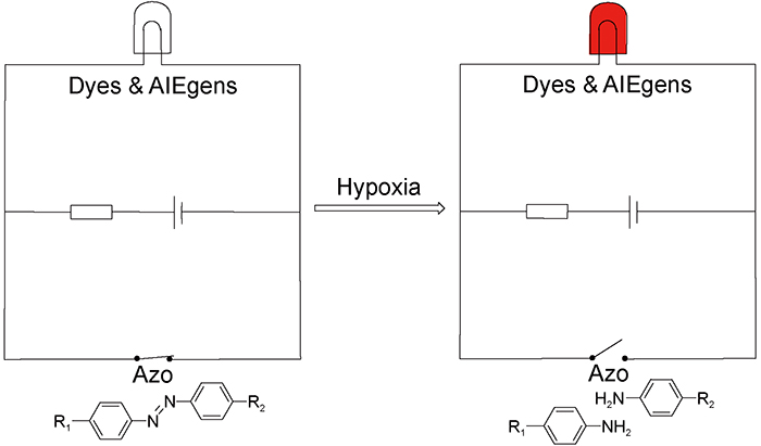
Application of Azobenzene Derivative Probes in Hypoxia Cell Imaging

Yunxue Wu1, Hengyi Zhang1,*(), Yu Liu1   

  1. 1 College of Chemistry, Nankai University,Tianjin 300071, China
  • Received: Revised: Online: Published:
  • Contact: Hengyi Zhang
  • Supported by:
    the National Natural Science Foundation of China(21772100)
Richhtml ( 54 ) PDF ( 613 ) Cited
Export

EndNote

Ris

BibTeX

Tumor tissues have lower oxygen concentration compared to normal tissues, due to the inadequate oxygen supply caused by uncontrolled cell growth and proliferation, in addition to an abnormal vasculature. As a common feature of solid tumors, hypoxia can be an indicator of malignant tissues or cancer progression. Accurate hypoxia detection and imaging are essential for the diagnosis and clinical treatment of cancer patients. Fluorescence imaging has been used in cancer detection because of its high sensitivity, non-invasive and real-time characteristics. During recent years, azo groups have been widely used to construct fluorescent probes for hypoxia cell imaging, owing to the fluorescence quenching effect on fluorophores and their reductive cleavage resulting in fluorescence recovery. This review summarizes various azobenzene derivative probes according to different construction strategies, and explores their mechanism and application in imaging. The limitations and future development of these probes are also discussed.

Contents

1 Introduction

2 Azobenzene derivative probes based on covalent strategies

2.1 Azobenzene derivative probes linked to dyes

2.2 Azobenzene derivative probes linked to AIEgens

3 Azobenzene derivative probes based on noncovalent strategies

4 Conclusion and outlook

Fig.1 Stepwise reduction of azobenzene derivatives to aniline derivatives[30]. Copyright 2010, American Chemical Society
Fig.2 (a) Design strategy of QCys.(b) Chemical structure of QCys[30]. Copyright 2010, American Chemical Society
Fig.3 Proposed mechanism of fluorescent probe HP for the detection of hypoxia[31]. Copyright 2015, The Royal Society of Chemistry
Fig.4 (a) Structures and detection mechanism of fluorescent probes MAR and MASR.(b) Fluorescence confocal microscopy images of MAR or MASR in live A549 cells[36]. Copyright 2013, Wiley-VCH Verlag GmbH & Co. KGaA, Weinheim
Fig.5 Fabrication of TPE-containing fluorescent polymeric aggregates and hypoxia detection mechanism[50]. Copyright 2018, American Chemical Society
Fig.6 Microscopy images of adherent A549 cells treated with PEG-Azo-TPE probes under normoxia(20% O2) and hypoxia(1% O2)[52]. Copyright 2019, Wiley-VCH Verlag GmbH & Co. KGaA, Weinheim
Fig.7 (a) Schematic illustration of the conventional covalent hypoxia-responsive probes.(b) Hypoxia detection mechanism of fluorescent probe CAC4A-Rho123.(c) Confocal laser scanning microscopy images of A549 cells incubated with CAC4A-Rho123 under hypoxic(less than 0.1% O2) or normoxic(20% O2) conditions for 8 h[62]. Copyright 2019, Wiley-VCH Verlag GmbH & Co. KGaA, Weinheim
Fig.8 Schematic illustration of polymersomes releasing dyes/drugs under hypoxia[64]. Copyright 2016, American Chemical Society
Fig.9 Fabrication of activatable polymeric AIE aggregates via selfassembly in an aqueous solution[66]. Copyright 2020, The Royal Society of Chemistry
[1]
Gilkes D M, Semenza G L, Wirtz D. Nat. Rev. Cancer, 2014, 14(6):430.
[2]
Vaupel P. Oncol., 2008, 13(S3):21.
[3]
Valastyan S, Weinberg R A. Cell, 2011, 147(2):275.
[4]
Bray F, Ferlay J, Soerjomataram I, Siegel R L, Torre L A, Jemal A. CA: A Cancer J. Clin., 2018, 68(6):394.
[5]
Torre L A, Bray F, Siegel R L, Ferlay J, Lortet-Tieulent J, Jemal A. CA: A Cancer J. Clin., 2015, 65(2):87.
[6]
Bach P B, Cramer L D, Warren J L, Begg C B. N Engl J. Med., 1999, 341(16):1198.
[7]
Zhang Z, Bast R C, Yu Y H, Li J N, Sokoll L J, Rai A J, Rosenzweig J M, Cameron B, Wang Y Y, Meng X Y, Berchuck A, van Haaften-Day C, Hacker N F, de Bruijn H W A, van der Zee A G J, Jacobs I J, Fung E T, Chan D W. Cancer Res., 2004, 64(16):5882.
[8]
Brown J M, Wilson W R. Nat. Rev. Cancer, 2004, 4(6):437.
[9]
Muller M, Padberg W, Schindler E, Sticher J, Osmer C, Friemann S, Hempelmann G. Anesth. Analg., 1998, 87(2):474.
[10]
CervÓs-Navarro J, Diemer N H. Crit. Rev. Neurobiol., 1991, 6:149.
[11]
Vaupel P, Schlenger K, Knoop C, Hçckel M. Cancer Res., 1991, 51:3316.
[12]
Parks S K, Cormerais Y, PouyssÉgur J. J. Physiol., 2017, 595(8):2439.
[13]
Schito L, Semenza G L. Trends Cancer, 2016, 2(12):758.
[14]
Eltzschig H K, Carmeliet P. N Engl J. Med., 2011, 364(7):656.
[15]
Minn H, Gronroos T, Komar G, Eskola O, Lehtio K, Tuomela J, Seppanen M, Solin O. Curr. Pharm. Des., 2008, 14(28):2932.
[16]
Jubb A M, Buffa F M, Harris A L. J. Cell. Mol. Med., 2010, 14(1/2):18.
[17]
Rajendran J G, Hendrickson K R G, Spence A M, Muzi M, Krohn K A, Mankoff D A. Eur. J. Nucl. Med. Mol. Imaging, 2006, 33(S1):44.
[18]
Chan J, Dodani S C, Chang C J. Nat. Chem., 2012, 4(12):973.
[19]
Zipfel W R, Williams R M, Webb W W. Nat. Biotechnol., 2003, 21(11):1369.
[20]
Stephens D J. Science, 2003, 300(5616):82.
[21]
Liu J N, Bu W B, Shi J L. Chem. Rev., 2017, 117(9):6160.
[22]
Cui L, Zhong Y, Zhu W P, Xu Y F, Du Q S, Wang X, Qian X H, Xiao Y. Org. Lett., 2011, 13(5):928.
[23]
Guo T, Cui L, Shen J N, Zhu W P, Xu Y F, Qian X H. Chem. Commun., 2013, 49(92):10820.
[24]
Li Z, He X, Wang Z, Yang R, Shi W, Ma H. Biosens. Bioelectron., 2015, 63:112.
[25]
Zhang J, Liu H W, Hu X X, Li J, Liang L H, Zhang X B, Tan W H. Anal. Chem., 2015, 87(23):11832.
[26]
Zhou J, Shi W, Li L H, Gong Q Y, Wu X F, Li X H, Ma H M. Chem. Asian J., 2016, 11(19):2719.
[27]
Zhang P Y, Huang H Y, Chen Y, Wang J Q, Ji L N, Chao H. Biomaterials, 2015, 53:522.
[28]
Komatsu H, Shindo Y, Oka K, Hill J P, Ariga K. Angew. Chem. Int. Ed., 2014, 53(15):3993.
[29]
Zbaida S, Levine W G. Chem. Res. Toxicol., 1991, 4(1):82.
[30]
Kiyose K, Hanaoka K, Oushiki D, Nakamura T, Kajimura M, Suematsu M, Nishimatsu H, Yamane T, Terai T, Hirata Y, Nagano T. J. Am. Chem. Soc., 2010, 132(45):15846.
[31]
Cai Q, Yu T, Zhu W P, Xu Y F, Qian X H. Chem. Commun., 2015, 51(79):14739.
[32]
Ti Y Z, Yu L, Tang Y, Jin T X, Yang M, Wang R, Xu Y F, Zhu W P. Sensor Actuat. B: Chem., 2018, 265:582.
[33]
Tang J R, Huang C X, Shu J Y, Zheng J, Ma D D, Li J S, Yang R H. Anal. Chem., 2018, 90(9):5865.
[34]
Liu N, Zou Z, Liu J, Zhu C, Zheng J, Yang R H. Anal., 2019, 144(21):6254.
[35]
Zhou F, Fu T, Huang Q, Kuai H L, Mo L T, Liu H L, Wang Q Q, Peng Y B, Han D M, Zhao Z L, Fang X H, Tan W H. J. Am. Chem. Soc., 2019, 141(46):18421.
[36]
Piao W, Nagano T, Hanaoka K. Angew. Chem. Int. Ed., 2013, 52:13028.
[37]
Shin N, Hanaoka K, Piao W, Miyakawa T, Fujisawa T, Takeuchi S, Takahashi S, Komatsu T, Ueno T, Terai T, Tahara T, Tanokura M, Nagano T, Urano Y. ACS Chem. Biol., 2017, 12(2):558.
[38]
Chevalier A, Piao W, Hanaoka K, Nagano T, Renard P Y, Romieu A. Methods Appl. Fluoresc., 2015, 3(4):044004.
[39]
Cui L, Shi Y P, Zhang S P, Yan L L, Zhang H, Tian Z R, Gu Y Y, Guo T, Huang J H. Dye. Pigment., 2017, 139:587.
[40]
Verwilst P, Han J Y, Lee J, Mun S, Kang H G, Kim J S. Biomaterials, 2017, 115:104.
[41]
Tian X W, Li Z, Sun Y, Wang P, Ma H M. Anal. Chem., 2018, 90(22):13759.
[42]
Luo J D, Xie Z L, Lam J W Y, Cheng L, Tang B Z, Chen H Y, Qiu C F, Kwok H S, Zhan X W, Liu Y Q, Zhu D B. Chem. Commun., 2001,(18):1740.
[43]
Hong Y N, Lam J W Y, Tang B Z. Chem. Commun., 2009,(29):4332.
[44]
Li K T, Lin Y J, Lu C. Chem. Asian J., 2019, 14(6):715.
[45]
Zhan R Y, Pan Y T, Manghnani P N, Liu B. Macromol. Biosci., 2017, 17(5):1600433.
[46]
Feng G X, Liu B. Acc. Chem. Res., 2018, 51(6):1404.
[47]
Xu G P, Tang Y H, Ma Y Y, Xu A, Lin W Y. Spectrochimica Acta Part A: Mol. Biomol. Spectrosc., 2018, 188:197.
[48]
Xu C H, Zou H, Zhao Z, Zhang P F, Kwok R T K, Lam J W Y, Sung H H Y, Williams I D, Tang B Z. Adv. Funct. Mater., 2019, 29(34):1903278.
[49]
Yang B, Zhang X Y, Zhang X Q, Huang Z F, Wei Y, Tao L. Mater. Today, 2016, 19(5):284.
[50]
Li S, Wang J L, Shen J J, Wu B, He Y N. ACS Macro Lett., 2018, 7(4):437.
[51]
Yuan X J, Wang Z, Li L S, Yu J W, Wang Y Q, Li H K, Zhang J D, Zhang Z B, Zhou N C, Zhu X L. Mater. Chem. Front., 2019, 3(6):1097.
[52]
Xue T H, Jia X Q, Wang J L, Xiang J Y, Wang W, Du J J, He Y N. Chem. Eur. J., 2019, 25(41):9634.
[53]
Lehn J M. Pure Appl. Chem., 1978, 50(9/10):871.
[54]
Ma J C, Dougherty D A. Chem. Rev., 1997, 97(5):1303.
[55]
Smithrud D B, Sanford E M, Chao I, Ferguson S B, Carcanague D R, Evanseck J D, Houk K N, Diederich F. Pure Appl. Chem., 1990, 62(12):2227.
[56]
Hancock R D. J. Chem. Educ., 1992, 69(8):615.
[57]
Tian R, Wang H M, Niu R F, Ding D. J. Colloid Interface Sci., 2015, 453:15.
[58]
Chen X M, Zhang Y M, Liu Y. Supramol. Chem., 2016, 28(9/10):817.
[59]
Vögtle F. Supramoleculare Chemie. Teubner, Stuttgart, 1991.
[60]
Lehn J M. Angew. Chem. Int. Ed. Engl., 1988, 27(1):89.
[61]
Liu Y H, Liu Y. Progress in Chemistry, 2019, 31(11): 1528.
(刘耀华, 刘育. 化学进展, 2019, 31(11): 1528.)
[62]
Geng W C, Jia S R, Zheng Z, Li Z H, Ding D, Guo D S. Angew. Chem. Int. Ed., 2019, 58(8):2377.
[63]
Si X H, Ma S, Xu Y D, Zhang D W, Shen N, Yu H Y, Zhang Y, Song W T, Tang Z H, Chen X S. J. Control. Release, 2020, 320:83.
[64]
Kulkarni P, Haldar M K, You S, Choi Y, Mallik S. Biomacromolecules, 2016, 17(8):2507.
[65]
Ma D D, Huang C X, Zheng J, Zhou W, Tang J R, Chen W J, Li J S, Yang R H. Anal. Chem., 2019, 91(2):1360.
[66]
Xue T H, Shao K C, Xiang J Y, Pan X Y, Zhu Z X, He Y N. Nanoscale, 2020, 12(14):7509.
[67]
Wang W L, Lin L, Ma X J, Wang B, Liu S R, Yan X X, Li S R, Tian H Y, Yu X F. ACS Appl. Mater. Interfaces, 2018, 10(23):19398.
[68]
Zhang X L, Wu M, Li J, Lan S Y, Zeng Y Y, Liu X L, Liu J F. ACS Appl. Mater. Interfaces, 2018, 10:21909.
[69]
Yan Q, Guo X L, Huang X L, Meng X, Liu F, Dai P P, Wang Z, Zhao Y J. ACS Appl. Mater. Interfaces, 2019, 11(27):24377.
[70]
Yang G B, Phua S Z F, Lim W Q, Zhang R, Feng L Z, Liu G F, Wu H W, Bindra A K, Jana D, Liu Z, Zhao Y L. Adv. Mater., 2019, 31(25):1901513.
[71]
Xu Z T, Pan C, Yuan W Z. Biomater. Sci., 2020, 8:3348.
[72]
Ihsanullah K M, Kumar B N, Zhao Y Y, Muhammad H, Liu Y, Wang L, Liu H, Jiang W. Biomaterials, 2020, 245:119982.
[73]
Lee J, Oh E T, Yoon H, Woo Kim C, Han Y J, Song J, Jang H, Joo Park H, Kim C. Nanoscale, 2017, 9(20):6901.
[74]
Li J J, Meng X, Deng J, Lu D, Zhang X, Chen Y R, Zhu J D, Fan A P, Ding D, Kong D L, Wang Z, Zhao Y J. ACS Appl. Mater. Interfaces, 2018, 10(20):17117.
[75]
Li S Y, Jiang X Y, Zheng R R, Zuo S J, Zhao L P, Fan G L, Fan J H, Liao Y H, Yu X Y, Cheng H. Chem. Commun., 2018, 54(57):7983.
[76]
Huang C X, Tan W L, Zheng J, Zhu C, Huo J, Yang R H. ACS Appl. Mater. Interfaces, 2019, 11(29):25740.
[1] Dang Zhang, Xi Wang, Lei Wang. Biomedical Applications of Enzyme-Powered Micro/Nanomotors [J]. Progress in Chemistry, 2022, 34(9): 2035-2050.
[2] Yanqin Lai, Zhenda Xie, Manlin Fu, Xuan Chen, Qi Zhou, Jin-Feng Hu. Construction and Application of 1,8-Naphthalimide-Based Multi-Analyte Fluorescent Probes [J]. Progress in Chemistry, 2022, 34(9): 2024-2034.
[3] Zilin Zhu, Zhongxian Fan, Mengzhao Miao, Huaiyi Huang. Photodynamic Therapy of Hypoxic Tumors with Ir(Ⅲ) Complexes [J]. Progress in Chemistry, 2021, 33(9): 1473-1481.
[4] Xiaodong Jing, Ying Sun, Bing Yu, Youqing Shen, Hao Hu, Hailin Cong. Rational Design of Tumor Microenvironment Responsive Drug Delivery Systems [J]. Progress in Chemistry, 2021, 33(6): 926-941.
[5] Jiajia Wang, Huiying Wu, Renfeng Dong, Yuepeng Cai. Micro/Nanomotors on the Way to Intelligent Cancer Diagnosis, Delivery and Therapy [J]. Progress in Chemistry, 2021, 33(5): 883-894.
[6] Yuanyuan Liu, Yun Guo, Xiaogang Luo, Genyan Liu, Qi Sun. Detection of Metal Ions, Small Molecules and Large Molecules by Near-Infrared Fluorescent Probes [J]. Progress in Chemistry, 2021, 33(2): 199-215.
[7] Shan Guo, Xiang Zhou. Detection of Circulating Tumor Cell in Vivo:Technology and Application [J]. Progress in Chemistry, 2021, 33(1): 1-12.
[8] Yunxue Xu, Renfu Liu, Kun xu, Zhifei Dai. Fluorescent Probes for Intraoperative Navigation [J]. Progress in Chemistry, 2021, 33(1): 52-65.
[9] Ziru Sun, Shengnan Liu, Qingzhi Gao. Development of Anticancer Drugs Targeting Glucose Transporters(GLUTs) [J]. Progress in Chemistry, 2020, 32(12): 1869-1878.
[10] Yue Li, Jinghong Li. CRISPR Bioanalytical Chemistry Technology [J]. Progress in Chemistry, 2020, 32(1): 5-13.
[11] Xinyi Lai, Zhiyong Wang, Yongtai Zheng, Yongming Chen. Nanoscale Metal Organic Frameworks for Drug Delivery [J]. Progress in Chemistry, 2019, 31(6): 783-790.
[12] Bingde Zheng, Yuanyuan Zhao, Hongcai Li, Biyuan Zheng, Meirong Ke, Jiandong Huang*. Activatable Photodynamic Anticancer Photosensitizers [J]. Progress in Chemistry, 2018, 30(9): 1403-1414.
[13] Liang He, Caiping Tan, Qian Cao, Zongwan Mao. Application of Phosphorescent Cyclometalated Iridium(Ⅲ) Complexes in Cancer Treatment [J]. Progress in Chemistry, 2018, 30(10): 1548-1556.
[14] Juan Shen, Yang Zhu, Hongdong Shi, Yangzhong Liu. Multifunctional Nanodrug Delivery Systems for Platinum-Based Anticancer Drugs [J]. Progress in Chemistry, 2018, 30(10): 1557-1572.
[15] Haidong Li, Jiangli Fan, Xiaojun Peng. Fluorescent Probes for the Recognition of Hypochlorous Acid [J]. Progress in Chemistry, 2017, 29(1): 17-35.