中文
Announcement
More
Progress in Chemistry 2021, Vol. 33 Issue (2): 216-231 DOI: 10.7536/PC20072 Previous Articles   Next Articles

• Review •

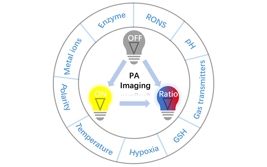
Applications of Activatable Organic Photoacoustic Contrast Agents

Jiawei Liu1, Jing Wang1, Qi Wang1,2,*(), Quli Fan1,*(), Wei Huang3   

  1. 1 Key Laboratory for Organic Electronics and Information Displays, Institute of Advanced Materials, Nanjing University of Posts and Telecommunications, Nanjing 210023, China
    2 State Key Laboratory of Bioelectronics, Southeast University, Nanjing 210096, China
    3 Institute of Flexible Electronics, Northwestern Polytechnical University, Xi’an 710072, China
  • Received: Revised: Online: Published:
  • Contact: Qi Wang, Quli Fan
  • About author:
    * Corresponding author e-mail: (Qi Wang);
  • Supported by:
    National Natural Science Foundation of China(21602112); National Natural Science Foundation of China(21674048); Open Research Fund of State Key Laboratory of Bioelectronics, Southeast University(OPSKLB202006); Postgraduate Research & Practice Innovation Program of Jiangsu Province(KYCX20_0752)
Richhtml ( 7 ) PDF ( 175 ) Cited
Export

EndNote

Ris

BibTeX

Photoacoustic(PA) imaging, as a new type of imaging technique that offers strong optical absorption contrast and high ultrasonic resolution, shows great application prospects in the early disease diagnosis for its characteristics of deep tissue penetration and high spatial resolution. However, traditional “always on” PA contrast agents have many disadvantages such as low signal-to-noise ratio, poor selectivity and specificity. In contrast, activatable PA contrast agents, where the imaging signal can be changed in response to pathologic parameters, have shown decreased background signal and improved selectivity and specificity in early disease diagnosis. Moreover, these contrast agents can obtain pathological parameters and information of various diseases at the molecular level by rational design to their structures, providing important guidelines for the optimization of treatment options. Therefore, activatable PA contrast agents hold greater promise in clinical practice than traditional “always on” PA contrast agents. In this review, we describe the recent advances in the development of activatable PA contrast agents. The design mechanisms and proof-of-concept applications of these activatable PA contrast agents are summarized in detail. The use of these activatable probes to detect different pathologic parameters(such as metal ions, enzymes, reactive nitrogen and reactive oxygen species) is highlighted. Finally, current challenges and future perspectives in this emerging field are also analyzed.

Contents

1 Introduction

2 “Always on” versus activatable PA imaging

3 Applications of activatable PA imaging contrast agents

3.1 Metal ions

3.2 Enzymes

3.3 RONS

3.4 pH

3.5 Gasotransmitters

3.6 Glutathione(GSH)

3.7 Hypoxia

3.8 Others

4 Conclusion and outlook

Fig. 1 Comparison of activatable PA probe and convetional PA probe and its related applications
Fig. 2 (A) Schematic illustration of PA probe LET-2 for the detection of Cu2+;(B) The absorption spectra of LET-2 upon treatment with Cu2+ and plot of PA715 against the concentration of Cu2+.[33] Copyright 2019, John Wiley and Sons
Fig. 3 (A) Schematic diagram of the probe NRh-IR-NMs for detection of Cu2+ and absorption spectra changes of the nanoprobe.[31] Copyright 2019, American Chemical Society;(B) Chemical structure of the APC-2 and the products formed after Cu(II) treatment;[32](C) Probe hCy7 and its reaction product hCy7'[34]
Fig. 4 (A) Schematic illustration of mechanism of Li+ probe reported by Clark et al.[37] Copyright 2015, American Chemical Society;(B) Absorption spectra of probe reported by Kopelman et al. at different concentrations of potassium(from 10 μmol/L to 1 mol/L). [40] Copyright 2017, American Chemical Society
Fig. 5 (A) Schematic illustration of the structure of the Probe 1 and it responsively self-assembled into nanofibers in tumor sites;(B) PA signal enhancing after 1 h co-incubation of gelatinase(15 ng·mL-1) and Probe 1;(C) The PA signal intensity in tumor site with time increase from 0.5 to 24 h postinjection.[45] Copyright 2015, John Wiley and Sons
Fig. 6 (A) Schematic illustration of condensation reactions and self-assembly of probe ESOR-PA01 at tumor microenvironment;(B) The PA imaging of the probe in Furin-overexpressing MDA-MB-231 cells and furin-deficient LoVo cells;(C) Photographic image of a mouse placed in a positioning device for PA imaging and representative PA images of mice tumors[48] Copyright 2013, American Chemical Society
Fig. 7 (A) The structure of probe P-Dex and its reaction product CyN3OH-Dex;(B) UV-Vis absorption and PA spectra of P-Dex or SP in the absence or presence of uPA.[50] Copyright 2020, John Wiley and Sons
Fig. 8 Design and characterization of the probe PCBP[61].(A) The activation and self-assembly mechanism of probe;(B) Representative PA intensity projection images of tumors after systemic administration of PCBP through tail vein. Copyright 2017, John Wiley and Sons
Fig. 9 (A) The oxidation reaction of ABTS and H2O2 in the probe Lipo@HRP&ABTS;[62](B) Schematic illustration of redox mechanism of probe BDP-DOH[63]
Fig. 10 (A) Preparation of probe PDI-IR790s-Fe/Pt;(B) Representative PA images at 680 and 790 nm of a subcutaneous U87MG tumor after intravenous injection and the PA signal intensity as a function of postinjection time.[67] Copyright 2018, John Wiley and Sons
Fig. 11 (A) Oxidation of molecule BBD in probe OSN-B1;[70](B) Activated mechanism of probe OEG-Aza-BODIPY-BAPE[71]
Fig. 12 (A) Protonation and deprotonation of IR-pH in probe LET-4;[74](B) Molecular structures of F-DTS and pH-BDP in Probe 3[77]
Fig. 13 (A) The structure of NP1 and the cleavage of hydrazone bonds in a physiologically acidic environment;(B) PA signal amplitude intensity at the two wavelengths of NP1 at different pH values and PA signal ratios between 785 nm and 708 nm.[79] Copyright 2012, Royal Society of Chemistry
Fig. 14 (A) Schematic illustration of the preparation of probe PBP@MnO2 NPs and its activation mechanism by H2O2/pH;(B) Representative PA images of 4T1 neoplastic mice after tail vein administration and ratiometric PA signals(ΔPA825/ΔPA680) as a function of the postinjection time[81] Copyright 2019, Royal Society of Chemistry
Fig. 15 (A) Detection mechanism of PA probe HS-CyBz;[83](B) Structures of CyCl-1 and CyCl-2 and the proposed mechanism for H2S detection;[84](C) Proposed mechanism for ratiometric photoacoustic detection of H2S by probe AzHD-LP[85]
Fig. 16 (A) The illustration of the reaction for NRM to NO in the probe DATN;[88](B) Response mechanism of probe APNO-5[89]
Fig. 17 (A) The disulfide bond of IR806-PDA can be cleaved by GSH and a subsequent exchange between —SH and the secondary amine occurred to form the thiolate-substituted IR806(IR806-S-NH 2);(B) PA signal amplitude intensity at 820 nm and 680 nm of IR806-PDA after injection and PA signal ratios between the two wavelengths.[92] Copyright 2018, John Wiley and Sons
Fig. 18 (A) Reduction process of HyP-1 in hypoxic environment;[94](B) Schematic illustration for the probe NR-azo response toward hypoxia[95]
Table 1 Summary of properties of partially activatable PA probes in this review
[1]
He S Q, Song J, Qu J L, Cheng Z. Chem. Soc. Rev., 2018, 47:4258.

pmid: 29725670
[2]
Hong G S, Antaris A L, Dai H J. Nat. Biomed. Eng., 2017, 1:0010.
[3]
Wang Q, Xu J Z, Geng R Y, Cai J, Li J, Xie C, Tang W H, Shen Q M, Huang W, Fan Q L. Biomaterials, 2020, 231:119671.
[4]
Weber J, Beard P C, Bohndiek S E. Nat. Methods, 2016, 13:639.
[5]
Lemaster J E, Jokerst J V. Wiley Interdiscip. Rev.: Nanomed. Nanobiotechnol., 2017, 9(1):11.
[6]
Upputuri P K, Pramanik M. J. Biomed. Opt., 2019, 24:1.
[7]
Jiang Y Y, Pu K Y. Small, 2017, 13:1700710.
[8]
Wang Q, Dai Y N, Xu J Z, Cai J, Niu X R, Zhang L, Chen R F, Shen Q M, Huang W, Fan Q L. Adv. Funct. Mater., 2019, 29:1901480.
[9]
Wang Q, Xia B, Xu J Z, Niu X R, Cai J, Shen Q M, Wang W J, Huang W, Fan Q L. Mater. Chem. Front., 2019, 3:650.
[10]
Ye J M, Li Z, Fu Q R, Li Q Q, Zhang X, Su L C, Yang H H, Song J B. Adv. Funct. Mater., 2020, 30:2001771.
[11]
Garcea S C, Wang Y, Withers P J. Compos. Sci. Technol., 2018, 156:305.
[12]
Parada S A, Eichinger J K, Dumont G D, Parada C A, Greenhouse A R, Provencher M T, Higgins L D, Warner J J P. Arthrosc.: J. Arthrosc. Relat. Surg., 2018, 34:84.
[13]
Poelma C. Exp. Fluids, 2017, 58:3.
[14]
Huang T J, Kim K T, Nakamura H, Yeung A T, Zeng J C. Biomed Res. Int., 2017, 2017:1.
[15]
Renard D. Eur. J. Neurol., 2018, 25:441.

doi: 10.1111/ene.13544 pmid: 29222944
[16]
Steinberg I, Huland D M, Vermesh O, Frostig H E, Tummers W S, Gambhir S S. Photoacoustic clinical imaging. Photoacoustics, 2019, 14:77.
[17]
Liu Y Y, Fan X, Li Y Y, Qu L L, Qin H Y, Cao Y N, Li H T. Prog. Chem., 2015, 27:1459.
刘迎亚, 范霄, 李艳艳, 渠陆陆, 覃海月, 曹英男, 李海涛. 化学进展, 2015, 27:1459.
[18]
Xu M H, Wang L V. Rev. Sci. Instruments, 2006, 77:041101.
[19]
Lu X M, Chen P F, Hu W B, Tang Y F, Huang W, Fan Q L. Prog. Chem., 2017, 29:119.
卢晓梅, 陈鹏飞, 胡文博, 唐玉富, 黄维, 范曲立. 化学进展, 2017, 29:119.
[20]
Wu J J, Yang Zhen, Jiao J M, Sun P F, Fan Q L, Huang W. Progress in Chemistry, 2017, 029(2):216.
吴锦钧, 杨震, 焦剑梅, 孙鹏飞, 范曲立, 黄维. 化学进展, 2017, 029(2):216.
[21]
Chen C, Ou H L, Liu R H, Ding D. Adv. Mater., 2020, 32:1806331.
[22]
Ou H L, Li J, Chen C, Gao H Q, Xue X, Ding D. Sci. China Mater., 2019, 62:1740.
[23]
Tang Y F, Pei F, Lu X M, Fan Q L, Huang W. Adv. Optical Mater., 2019, 7:1900917.
[24]
Wang X H, Wang X Y, Jin S X, Muhammad N, Guo Z J. Chem. Rev., 2019, 119:1138.

pmid: 30299085
[25]
Miao Q Q, Pu K Y. Bioconjugate Chem., 2016, 27:2808.
[26]
Lyu Y, Pu K Y. Adv. Sci., 2017, 4:1600481.
[27]
Fu Q, Zhu R, Song J, Yang H, Chen X. Adv. Mater., 2019, 31(6):e1805875.

pmid: 30556205
[28]
Carter K P, Young A M, Palmer A E. Chem. Rev., 2014, 114:4564.
[29]
Knox H J, Chan J. Acc. Chem. Res., 2018, 51:2897.

doi: 10.1021/acs.accounts.8b00351 pmid: 30379532
[30]
Wang S C, Sheng Z H, Yang Z G, Hu D H, Long X J, Feng G, Liu Y B, Yuan Z, Zhang J J, Zheng H R, Zhang X J. Angew. Chem. Int. Ed., 2019, 58:12415.
[31]
Wang W, Tran R, Qu J F, Liu Y, Chen C, Xu M G, Chen Y B, Ong S P, Wang L Z, Zhou W, Shao Z P. ACS Appl. Mater. Interfaces, 2019, 11:35641.

doi: 10.1021/acsami.9b07966 pmid: 31532199
[32]
Li H, Zhang P, Smaga L P, Hoffman R A, Chan J. J. Am. Chem. Soc., 2015, 137:15628.
[33]
Zeng L L, Ma G C, Xu H, Mu J, Li F, Gao X T, Deng Z T, Qu J L, Huang P, Lin J. Small, 2019, 15:1803866.
[34]
Liu Y, Wang S, Ma Y, Lin J, Wang H Y, Gu Y Q, Chen X Y, Huang P. Adv. Mater., 2017, 29:1606129.
[35]
Huang Y, Li F, Ma G C, Yang W W, Zhang X F, Lin J, Luo Y X, Huang P. Talanta, 2018, 187:65.
[36]
Roberts S, Seeger M, Jiang Y Y, Mishra A, Sigmund F, Stelzl A, Lauri A, Symvoulidis P, Rolbieski H, Preller M, Deán-Ben X L, Razansky D, Orschmann T, Desbordes S C, Vetschera P, Bach T, Ntziachristos V, Westmeyer G G. J. Am. Chem. Soc., 2018, 140:2718.
[37]
Cash K J, Li C Y, Xia J, Wang L V, Clark H A. ACS Nano, 2015, 9:1692.

pmid: 25588028
[38]
Ho I T, Sessler J L, Gambhir S S, Jokerst J V. Anal., 2015, 140:3731.
[39]
Xiang J J, Liu C J, Zhou L H, Yang X, Li Y, Jiang Y C, Mahmood T, Zhang P F, Gong P, Cai L T. Anal. Chem., 2020, 92:4721.
[40]
Lee C H, Folz J, Zhang W L, Jo J, Tan J W Y, Wang X D, Kopelman R. Anal. Chem., 2017, 89:7943.
[41]
Mu J, Lin J, Huang P, Chen X Y. Chem. Soc. Rev., 2018, 47:5554.

doi: 10.1039/C7CS00663B
[42]
Razgulin A, Ma N, Rao J H. Chem. Soc. Rev., 2011, 40:4186.

pmid: 21552609
[43]
Zhang J D, Liu H Z, Meng L. Chinese Journal of Organic Chemistry, 2019, 39(11):3132.
张继东, 刘鸿泽, 孟丽. 有机化学, 2019, 39(11):3132.
[44]
Wu C F, Zhang R, Du W, Cheng L, Liang G L. Nano Lett., 2018, 18:7749.
[45]
Zhang D, Qi G B, Zhao Y X, Qiao S L, Yang C, Wang H. Adv. Mater., 2015, 27:6125.
[46]
Li L L, Ma H L, Qi G B, Zhang D, Yu F Q, Hu Z Y, Wang H. Adv. Mater., 2016, 28:254.

pmid: 26568542
[47]
Lin Y X, Wang Y, Qiao S L, An H W, Wang J, Ma Y, Wang L, Wang H. Biomaterials, 2017, 141:199.
[48]
Dragulescu-Andrasi A, Kothapalli S R, Tikhomirov G A, Rao J H, Gambhir S S. J. Am. Chem. Soc., 2013, 135:11015.
[49]
Wang Y Q, Hu X M, Weng J H, Li J B, Fan Q L, Zhang Y, Ye D J. Angew. Chem. Int. Ed., 2019, 58:4886.
[50]
Li Q, Li S H, He S S, Chen W, Cheng P H, Zhang Y, Miao Q Q, Pu K Y. Angew. Chem. Int. Ed., 2020, 59:7018.
[51]
Gao X T, Ma G C, Jiang C, Zeng L L, Jiang S S, Huang P, Lin J. Anal. Chem., 2019, 91:7112.
[52]
Levi J, Kothapalli S R, Bohndiek S, Yoon J K, Dragulescu-Andrasi A, Nielsen C, Tisma A, Bodapati S, Gowrishankar G, Yan X R, Chan C, Starcevic D, Gambhir S S. Clin. Cancer Res., 2013, 19:1494.

doi: 10.1158/1078-0432.CCR-12-3061
[53]
Zhang J J, Smaga L P, Satyavolu N S R, Chan J, Lu Y. J. Am. Chem. Soc., 2017, 139:17225.

pmid: 29028325
[54]
Meng X Q, Zhang J L, Sun Z H, Zhou L H, Deng G J, Li S P, Li W J, Gong P, Cai L T. Theranostics, 2018, 8:6025.
[55]
Bauerová K, Bezek A. Gen. Physiol. Biophys., 1999, 18:15.

pmid: 10703714
[56]
Martin L D, Krunkosky T M, Dye J A, Fischer B M, Jiang N F, Rochelle L G, Akley N J, Dreher K L, Adler K B. Environ. Heal. Perspect., 1997, 105:1301.
[57]
Baran C, Zeigler M, Tridandapani S, Marsh C. Curr. Pharm. Des., 2004, 10:855.
[58]
Tapeinos C, Pandit A. Adv. Mater., 2016, 28:5553.
[59]
Ye H, Zhou Y, Liu X, Chen Y B, Duan S Z, Zhu R Y, Liu Y, Yin L C. Biomacromolecules, 2019, 20:2441.

pmid: 31117357
[60]
Jiao C P, Liu Y Y, Lu W J, Zhang P P, Wang Y F. Chinese Journal of Organic Chemistry, 2019, 39(3):591.
矫春鹏, 刘媛媛, 路文娟, 张平平, 王延风. 有机化学, 2019, 39(3):591.
[61]
Xie C, Zhen X, Lyu Y, Pu K Y. Adv. Mater., 2017, 29:1703693.
[62]
Chen Q, Liang C, Sun X Q, Chen J W, Yang Z J, Zhao H, Feng L Z, Liu Z. PNAS, 2017, 114:5343.

pmid: 28484000
[63]
Zheng J D, Zeng Q, Zhang R J, Xing D, Zhang T. J. Am. Chem. Soc., 2019, 141:19226.

doi: 10.1021/jacs.9b10353 pmid: 31770490
[64]
Wang Z M, Zhen X, Upputuri P K, Jiang Y Y, Lau J, Pramanik M, Pu K Y, Xing B G. ACS Nano, 2019, 13:5816.
[65]
Yang Y L, Wang S F, Lu L F, Zhang Q S, Yu P, Fan Y, Zhang F. Angew. Chem. Int. Ed., 2020, 59:18380.
[66]
Yin C, Zhen X, Fan Q L, Huang W, Pu K Y. ACS Nano, 2017, 11:4174.

pmid: 28296388
[67]
Yang Z, Dai Y L, Yin C, Fan Q L, Zhang W S, Song J, Yu G C, Tang W, Fan W P, Yung B C, Li J, Li X, Li X C, Tang Y F, Huang W, Song J B, Chen X Y. Adv. Mater., 2018, 30:1707509.
[68]
Pu K Y, Shuhendler A J, Jokerst J V, Mei J G, Gambhir S S, Bao Z N, Rao J H. Nat. Nanotechnol., 2014, 9:233.
[69]
Qin X L, Li F, Zhang Y F, Ma G C, Feng T, Luo Y X, Huang P, Lin J. Anal. Chem., 2018, 90:9381.

doi: 10.1021/acs.analchem.8b01992 pmid: 29963863
[70]
Zhang J J, Zhen X, Upputuri P K, Pramanik M, Chen P, Pu K Y. Adv. Mater., 2017, 29:1604764.
[71]
Lu X M, Zhao M, Chen P F, Fan Q L, Wang W J, Huang W. J. Mater. Chem. B, 2018, 6:4531.

pmid: 32254670
[72]
Toriumi N, Asano N, Ikeno T, Muranaka A, Hanaoka K, Urano Y, Uchiyama M. Angew. Chem. Int. Ed., 2019, 58:7788.
[73]
Adrogué H J, Madias N E. N Engl J. Med., 1998, 338:107.
[74]
Ma G C, Gao X T, Jiang C, Xing S J, Wei C L, Huang P, Lin J. Anal. Chem., 2019, 91:13570.

doi: 10.1021/acs.analchem.9b02701 pmid: 31610654
[75]
Chen Q, Liu X D, Zeng J F, Cheng Z P, Liu Z. Biomaterials, 2016, 98:23.

pmid: 27177219
[76]
Guha S, Shaw G K, Mitcham T M, Bouchard R R, Smith B D. Chem. Commun., 2016, 52:120.
[77]
Miao Q Q, Lyu Y, Ding D, Pu K Y. Adv. Mater., 2016, 28:3662.

pmid: 27000431
[78]
Chen Q, Liu X D, Chen J W, Zeng J F, Cheng Z P, Liu Z. Adv. Mater., 2015, 27:6820.
[79]
Huang G J, Si Z, Yang S H, Li C, Xing D. J. Mater. Chem., 2012, 22:22575.
[80]
Ju K Y, Kang J, Pyo J, Lim J, Chang J H, Lee J K. Nanoscale, 2016, 8:14448.
[81]
Hu X M, Zhan C, Tang Y F, Lu F, Li Y Y, Pei F, Lu X M, Ji Y, Li J, Wang W J, Fan Q L, Huang W. Chem. Commun., 2019, 55:6006.
[82]
Wang R. Physiol. Rev., 2012, 92:791.

pmid: 22535897
[83]
Chen Z Y, Mu X L, Han Z, Yang S P, Zhang C L, Guo Z J, Bai Y, He W J. J. Am. Chem. Soc., 2019, 141:17973.

doi: 10.1021/jacs.9b09181 pmid: 31657918
[84]
Li X, Tang Y F, Li J, Hu X M, Yin C, Yang Z, Wang Q, Wu Z Z, Lu X M, Wang W J, Huang W, Fan Q L. Chem. Commun., 2019, 55:5934.
[85]
Ma T, Zheng J D, Zhang T, Xing D. Nanoscale, 2018, 10:13462.

pmid: 29972183
[86]
Denninger J W, Marletta M A. Biochim. et Biophys. Acta BBA - Bioenerg., 1999, 1411:334.
[87]
Wang L Z, Zhang J, An X, Duan H D. Org. Biomol. Chem., 2020, 18:1522.

pmid: 31995085
[88]
Teng L L, Song G S, Liu Y C, Han X Y, Li Z, Wang Y J, Huan S Y, Zhang X B, Tan W H. J. Am. Chem. Soc., 2019, 141:13572.

doi: 10.1021/jacs.9b05901 pmid: 31370392
[89]
Reinhardt C J, Zhou E Y, Jorgensen M D, Partipilo G, Chan J. J. Am. Chem. Soc., 2018, 140:1011.

pmid: 29313677
[90]
Kleinman W A, Richie J P Jr. Biochem. Pharmacol., 2000, 60:19.

doi: 10.1016/s0006-2952(00)00293-8 pmid: 10807941
[91]
Jung H S, Chen X Q, Kim J S, Yoon J. Chem. Soc. Rev., 2013, 42:6019.

pmid: 23689799
[92]
Yin C, Tang Y F, Li X Z, Yang Z, Li J, Li X, Huang W, Fan Q L. Small, 2018, 14:1703400.
[93]
Tang L G, Yu F, Tang B W, Yang Z, Fan W P, Zhang M R, Wang Z T, Jacobson O, Zhou Z J, Li L, Liu Y J, Kiesewetter D O, Tang W, He L C, Ma Y, Niu G, Zhang X Z, Chen X Y. ACS Appl. Mater. Interfaces, 2019, 11:27558.

pmid: 31317730
[94]
Knox H J, Hedhli J, Kim T W, Khalili K, Dobrucki L W, Chan J. Nat. Commun., 2017, 8:1794.

pmid: 29176550
[95]
Huang J, Wu Y L, Zeng F, Wu S Z. Theranostics, 2019, 9:7313.

doi: 10.7150/thno.36755 pmid: 31695770
[96]
Ng K K, Shakiba M, Huynh E, Weersink R A, Roxin Á, Wilson B C, Zheng G. ACS Nano, 2014, 8:8363.
[97]
Xiao H B, Wu C C, Li P, Gao W, Zhang W, Zhang W, Tong L L, Tang B. Chem. Sci., 2017, 8:7025.

doi: 10.1039/c7sc02330h pmid: 29147529
[1] Zhen Wang, Xi Li, Yuanyuan Li, Qi Wang, Xiaomei Lu, Quli Fan. Activatable NIR-Ⅱ Probe for Tumor Imaging [J]. Progress in Chemistry, 2022, 34(1): 198-206.
[2] Xiaodong Jing, Ying Sun, Bing Yu, Youqing Shen, Hao Hu, Hailin Cong. Rational Design of Tumor Microenvironment Responsive Drug Delivery Systems [J]. Progress in Chemistry, 2021, 33(6): 926-941.
[3] Fei Ren, Jianbing Shi, Bin Tong, Zhengxu Cai, Yuping Dong. Near Infrared Fluorescent Dyes with Aggregation-Induced Emission [J]. Progress in Chemistry, 2021, 33(3): 341-354.
[4] Pingping Zhao, Junxing Yang, Jianhui Shi, Jingyi Zhu. Construction and Application of Dendrimer-Based SPECT Imaging Agent [J]. Progress in Chemistry, 2021, 33(3): 394-405.
[5] Qing Wu, Yiyuan Tang, Miao Yu, Yueying Zhang, Xingmei Li. Stimuli-Responsive DNA Nanostructure Drug Delivery System Based on Tumor Microenvironment [J]. Progress in Chemistry, 2020, 32(7): 927-934.
[6] Jidong Zhang, Achen Liu, Jiao Chen, Guanghui Yuan, Huafeng Jin. Fluorescent Organic Small Molecule Based on Biotin and Their Applications [J]. Progress in Chemistry, 2020, 32(5): 594-603.
[7] Man Du, Baolong Huo, Jiemin Liu*, Mengwen Li, Leqiu Fang, Yunxu Yang*. Fluorescent Probes Based on Silicon-Substituted Xanthene Dyes and Their Applications in Bioimaging [J]. Progress in Chemistry, 2018, 30(6): 809-830.
[8] Guang Deng, Hong Yang, Zhiguo Zhou*, Shiping Yang*. Design and Application of T1-T2 Dual-Modal MRI Contrast Agents [J]. Progress in Chemistry, 2018, 30(10): 1534-1547.
[9] Xiaomei Lu, Pengfei Chen, Wenbo Hu, Yufu Tang, Wei Huang, Quli Fan. Organic Optoelectronic Materials for Photoacoustic Imaging [J]. Progress in Chemistry, 2017, 29(1): 119-126.
[10] Liu Yingya, Fan Xiao, Li Yanyan, Qu Lulu, Qin Haiyue, Cao Yingnan, Li Haitao. Multispectral Photoacoustic Tomography and Its Development in Biomedical Application [J]. Progress in Chemistry, 2015, 27(10): 1459-1469.
[11] Shen Aijun, Dong Haiqing, Wen Huiyun, Xu Meng, Li Yongyong, Wang Peijun. Gadolinium-Based Contrast Agents for Tumor Targeting Imaging [J]. Progress in Chemistry, 2011, 23(4): 772-780.
[12] Wang Lingyun Chen Qiuyun Wei Bin. MRI Contrast Agent for the Diagnosis of Tumor [J]. Progress in Chemistry, 2010, 22(01): 186-193.
[13]

Zhang Shanrong, Ren Jimin, Pei Fengkui

. The Research Progress of Magnetic Resonance Imaging Contrast Agents [J]. Progress in Chemistry, 1995, 7(02): 98-.