中文
Announcement
More
Progress in Chemistry 2020, Vol. 32 Issue (5): 656-664 DOI: 10.7536/PC190929 Previous Articles   Next Articles

• Review •

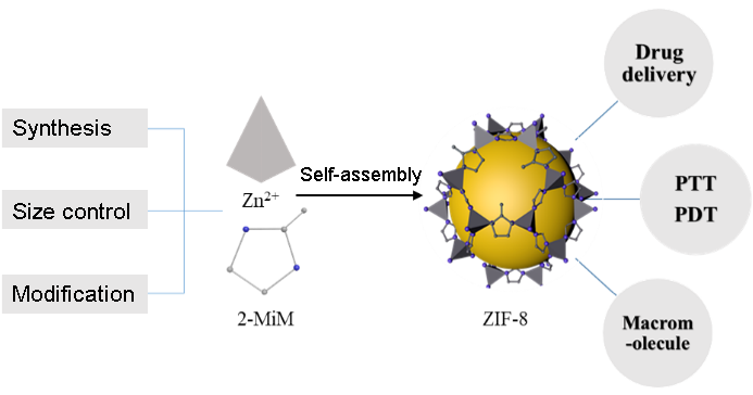
Size Control and Biomedical Applications of ZIF-8 Nanoparticles

Qiangqiang Hu1, Heze Guo1, Hongjing Dou1,**()   

  1. School of Materials Science and Engineering, State Key Laboratory of Metal Matrix Composites, Shanghai Jiao Tong University, Shanghai 200240, China
  • Received: Revised: Online: Published:
  • Contact: Hongjing Dou
  • About author:
  • Supported by:
    National Natural Science Foundation of China(21871180); Shanghai Science and Technology Innovation Action Plan Basic Research Area Project(18JC1413500); Shanghai University Distinguished Professor(Oriental Scholar) Program(SHDP201802); Shanghai Municipal Education Commission “Dawning Plan”(12SG12)
Richhtml ( 333 ) PDF ( 6452 ) Cited
Export

EndNote

Ris

BibTeX

Zeolitic imidazolate framework-8(ZIF-8) is a class of porous crystalline materials formed by the self-assembly of zinc ions and 2-methylimidazole. It shows potential advantages in encapsulation and transportation of functional materials owing to its high porosity, large specific surface,convenient synthesis and controllable size. More importantly, ZIF-8 is an ideal carrier for drug delivery and release due to its excellent biocompatibility, stability under physiological conditions and responsiveness to the weak acidic environment associated with malignant tumors and other diseases. In fact, the small molecular drugs(doxorubicin, 5-fluorouracil) and biological macromolecules(antibody, nuclein) have all been loaded into ZIF-8 for chemotherapy, photothermal therapy, photodynamic therapy and biosensing. The particle size of ZIF-8 is very important for biomedical applications, and accurate functional regulation of ZIF-8 is agreat challenge for its biomedical application. Herein, we review the synthesis methods, size-control and biomedicalapplications of ZIF-8.

Contents

1 Introduction

2 Synthesis of ZIF-8 nanoparticles

2.1 Solvothermal process

2.2 Microwave-assist

2.3 Microfluidic

3 Size control of ZIF-8

3.1 Formation mechanism

3.2 Size control

4 Biomedical applications

4.1 Application in tumor treatment

4.2 Application in bioimagings

4.3 Protective coating for biomacromolecule

5 Conclusion and outlook

Fig. 1 Surfactant-free synthesis by simply mixing precursors in appropriate solvents either at room temperature or at elevatedtemperature[41]. Copyright 2015, American Chemical Society
Fig. 2 Schematic illustration of the importance of nanosized PCP crystals with the enhanced contribution of crystal interfaces[5]. Copyright 2010, American Chemical Society
Fig. 3 Theoretical basis of the reaction-diffusion framework(RDF). (A) Diffusion profiles of HmIm(outer) and Zn2+/Co2+(inner) are depicted at a given time. Evolution of the reaction zone is also shown and exhibits a decrease of its amplitude and broadening of its width. Xf denotes thelocation of the peak which also corresponds to the location of the precipitation front.(B) Nucleation of pure ZIF-8 leading to nanosperoids takesplace within the reaction zone[19]. Copyright 2018, American Chemical Society
Fig. 4 A schematic illustration showing the influence of TEA concentration on ZIF-8 formation[27]. Copyright 2014, The Royal Society of Chemistry
Fig. 5 The pH-induced one-pot synthesis of MOFs with encapsulated target molecules[29]. Copyright 2015, American Chemical Society
Fig. 6 Possible mechanism for the dissociation of CuS@ZIF-8 NPs underthe laser irradiation (a). Cell viability of MCF-7 cells after 24 h incubation inthe presence of different concentrations of DOX or DOX loaded CuS@ZIF-8NPs(b). Cell viability of MCF-7 cells treated with PBS solution as a control,CuS@ZIF-8(25 mg·mL-1), DOX(1 mg·mL-1) or DOX loaded CuS@ZIF-8 NPs(1 mg·mL-1 of DOX, 4% DOX/NPs, w/w) for 12 h and then exposed to the NIR laser for 1, 2, 4, and 6 min(c). Photothermal images of mice, saline(control, group Ⅰ), DOX(group Ⅱ) and DOX loaded CuS@ZIF-8(group Ⅲ) under NIR laser irradiation for 3 min(d)[37]. Copyright 2016, The Royal Society of Chemistry
Fig. 7 Schematic illustration of Fe3O4@PAA/AuNCs/ZIF-8 composite NPs for simultaneous tri-modal cancer imagingand chemotherapy[55]. Copyright 2015, The Royal Society of Chemistry
Fig. 8 Characterization of biomimetically mineralized biocomposite. (a) SEM image showing the crystals obtained using BSA as a growth agentfor biomimetic mineralization(scale bar, 1 μm).(b) Photograph and(c) confocal laser scanning microscopy image of the biomomimetically mineralized ZIF-8 composite obtained using BSA labelled with FITC. This biocomposite(ZIF-8/FITC-BSA) was prepared at 37 ℃, washed and exposed to ultraviolet light of wavelength 365 and 495 nm, respectively(scale bar, 10 μm).(d) PXRD of the MOF-BSA biocomposite.(e) FTIR spectra of BSA(red), ZIF-8/BSA(orange), standard ZIF-8 post incubated with BSA after washing(blue), and standard ZIF-8(black).(f) SAXS data of the ZIF-8/BSA biocompositeand a schematic showing the relative size of BSA to the mesopore.(g) Schematic proposing the biomimetically mineralized growthof ZIF-8. Each BSA molecule attracts 31 2-methylimidazole(HmIm) ligands and 22 Zn2+ ions, facilitating the nucleation of ZIF-8 crystals[60]. Copyright 2015, Springer Nature
[1]
Echaide-G C, Clément C, Cacho-Bailo C, Téllez C, Coronas J. J. Mater. Chem. A, 2018,6:5485. http://xlink.rsc.org/?DOI=C8TA01232F

doi: 10.1039/C8TA01232F
[2]
Safaei M, Foroughi M M, Ebrahimpoor N, ShohrehJ,Omidi A, Khatami M. Trends in Analytical Chemistry, 2019,118:401. https://linkinghub.elsevier.com/retrieve/pii/S0165993619301840

doi: 10.1016/j.trac.2019.06.007
[3]
Hoopa M, Waldea C F, Riccòc R, Mushtaqa F, Terzopouloua A, Chena X Z, deMellob A J, Doonand C J, Falcaroc P, Nelsona B J, Puigmartí-Luisb J, PanéaaInstitute S. Applied Materials Today, 2018,11:13. https://linkinghub.elsevier.com/retrieve/pii/S2352940717303839

doi: 10.1016/j.apmt.2017.12.014
[4]
Tanaka S, KidaK,Okita M, Ito Y, Miyake Y. Chem. Lett, 2012,41:1337. http://www.journal.csj.jp/doi/10.1246/cl.2012.1337

doi: 10.1246/cl.2012.1337
[5]
Diring S, Furukawa S, Takashima Y, Tsuruoka T, Kitagawa S. Chem. Mater., 2010,22:4531. https://pubs.acs.org/doi/10.1021/cm101778g

doi: 10.1021/cm101778g
[6]
Chowdhuri A R, Das B, Kumar A, Tripathy S, Roy S, Kumar-Sahu S. Nanotecchnology, 2017,28:095102.
[7]
Chen Y, Tang S. J. Solid. State. Chem., 2019,276:68. https://linkinghub.elsevier.com/retrieve/pii/S0022459619302117

doi: 10.1016/j.jssc.2019.04.034
[8]
Rubio-Martinez M, Avci-Camur C, Thornton A W, Imaz I, Maspoch D, Hill M R. Chem. Soc. Rev., 2017,46:3453. http://xlink.rsc.org/?DOI=C7CS00109F

doi: 10.1039/C7CS00109F
[9]
Echaide-Górriz C, Clément C, Cacho-Bailo F, Téllez C, Coronas J. J. Mater. Chem. A, 2018,6:5485. http://xlink.rsc.org/?DOI=C8TA01232F

doi: 10.1039/C8TA01232F
[10]
Lee Y R, Jang M S, Cho H Y, Kwon H J, Kim S, Ahn W S. Chemical Engineering Journal, 2015,271:276. https://linkinghub.elsevier.com/retrieve/pii/S1385894715003034

doi: 10.1016/j.cej.2015.02.094
[11]
Cravillon J, Münzer S, Lohmeier S J, Feldhoff A, Huber K, Wiebcke M. Chem. Mater., 2009,21:1410. https://pubs.acs.org/doi/10.1021/cm900166h

doi: 10.1021/cm900166h
[12]
Pan Y P, Liu Y Y, Zeng G F, Zhao L, Lai Z P. Chem. Commun., 2011,47:2071. http://xlink.rsc.org/?DOI=c0cc05002d

doi: 10.1039/c0cc05002d
[13]
Khan N A, Jhung S H. Coord. Chem. Rev., 2015,285:11. https://linkinghub.elsevier.com/retrieve/pii/S0010854514002811

doi: 10.1016/j.ccr.2014.10.008
[14]
Butova V V, Budnik A P, Bulanova E A, Soldatov A V. Mendeleev Commun., 2016,26:43. https://linkinghub.elsevier.com/retrieve/pii/S0959943616000183

doi: 10.1016/j.mencom.2016.01.017
[15]
Awadallah-F A, Hillman F, Al-Muhtaseb S A, Jeong H K. Dalton Trans., 2019,48:4685. http://xlink.rsc.org/?DOI=C9DT00222G

doi: 10.1039/C9DT00222G
[16]
Wang X G, Cheng Q, Yu Y, Zhang X Z. Angew. Chem. Int. Ed., 2018,57:7836. http://doi.wiley.com/10.1002/anie.v57.26

doi: 10.1002/anie.v57.26
[17]
Yamamoto D, Maki T, Watanabe S, Tanaka H, Miyahara M T, Mae K. Chemical Engineering Journal, 2013,227:145. http://dx.doi.org/10.1016/j.cej.2012.08.065

doi: 10.1016/j.cej.2012.08.065
[18]
Van-Vleet M J, Weng T T, Li X Y, Schmidt J R. Chem. Rev. 2018,118:3681. https://pubs.acs.org/doi/10.1021/acs.chemrev.7b00582

doi: 10.1021/acs.chemrev.7b00582
[19]
Saliba D, Ammar M, Rammal M, Al-Ghoul M, Hmadeh M. J. Am. Chem. Soc., 2018,140:1812. https://pubs.acs.org/doi/10.1021/jacs.7b11589

doi: 10.1021/jacs.7b11589
[20]
Sosso G C, Chen J, Cox S J, Fitzner M, Pedevilla P, Zen A, Michaelides A. Chem. Rev., 2016,116:7078. https://pubs.acs.org/doi/10.1021/acs.chemrev.5b00744

doi: 10.1021/acs.chemrev.5b00744
[21]
Karthika S, Radhakrishnan T K, Kalaichelvi P A. Cryst. Growth Des., 2016,16:6663. https://pubs.acs.org/doi/10.1021/acs.cgd.6b00794

doi: 10.1021/acs.cgd.6b00794
[22]
Flügel E A, Ranft A, Haase F, Lotsch B V. J. Mater. Chem., 2012,22:10119. http://xlink.rsc.org/?DOI=c2jm15675j

doi: 10.1039/c2jm15675j
[23]
Rimer, J D, Tsapatsis, M J. MRS Bull., 2016,41:393. https://www.cambridge.org/core/product/identifier/S0883769416000890/type/journal_article

doi: 10.1557/mrs.2016.89
[24]
Beha J J, Lima J K, Ngb E P, Onia B S. Mater. Chem. Phys., 2018,216:393. https://linkinghub.elsevier.com/retrieve/pii/S0254058418305212

doi: 10.1016/j.matchemphys.2018.06.022
[25]
Zhang Y Y, Jia Y, Li M, Hou L A. Sci. Rep., 2018,8:8597. https://doi.org/10.1038/s41598-018-26926-z

doi: 10.1038/s41598-018-26926-z
[26]
Pan Y C, Heryadi D D, Zhou F, Zhao L, Lestari G, Sub H, Lai Z P. CrystEngComm., 2011,13:6937. http://dx.doi.org/10.1039/c1ce05780d

doi: 10.1039/c1ce05780d
[27]
Nordin N A H M, Ismail A F, Mustafa A, Goh P S, Rana D, Matsuura T. RSC Adv., 2014,4:33292. http://xlink.rsc.org/?DOI=C4RA03593C

doi: 10.1039/C4RA03593C
[28]
Cravillon J, Nayuk R, Springer S, Feldhoff A, Huber K, Wiebcke M. Chem. Mater., 2011,23:2130. https://pubs.acs.org/doi/10.1021/cm103571y

doi: 10.1021/cm103571y
[29]
Zheng H Q, Zhang Y N, Liu L F, Wan W, Guo P, Nystroöm A M, Zou X D. J. Am. Chem. Soc., 2016,138:962. https://pubs.acs.org/doi/10.1021/jacs.5b11720

doi: 10.1021/jacs.5b11720
[30]
Sun C Y, Qin C, Wang X L, Yang G S, Shao K Z, Lan Y Q, Su Z M, Huang P, Wang C G, Wang EB. Dalton Trans., 2012,41:6906. http://xlink.rsc.org/?DOI=c2dt30357d

doi: 10.1039/c2dt30357d
[31]
Zhang H Y, Li Q, Liu R L, Zhang X K, Li Z H, Luan Y X. Adv. Funct. Mater., 2018,28:1802830. http://doi.wiley.com/10.1002/adfm.v28.35

doi: 10.1002/adfm.v28.35
[32]
Wang H H, Li T, Li J W, Tong W J, Gao C Y. Colloids and Surfaces A, 2019,568:224. https://linkinghub.elsevier.com/retrieve/pii/S0927775719301128

doi: 10.1016/j.colsurfa.2019.02.025
[33]
Zou Q, Huang J N, Zhang X J. Small, 2018,14:1803101. http://doi.wiley.com/10.1002/smll.v14.45

doi: 10.1002/smll.v14.45
[34]
Zhao P H, Jin Z K, Chen Q, Yang T, Chen D Y, Meng Y, Lu X F, Gu Z, He Q J. Nat. Commun., 2018,9:1. http://www.nature.com/articles/s41467-017-02088-w

doi: 10.1038/s41467-017-02088-w
[35]
Wang W Q, Wang L, Li Y, Liu S, Xie Z G, Jing X B. Adv. Mater., 2016,28:9320. http://doi.wiley.com/10.1002/adma.201602997

doi: 10.1002/adma.201602997
[36]
Silva J S F, Silva J Y R, Sá G F, Araújo S S, Gomes-Filho M A, Ronconi C M, Santos CM, Júnior S. A. ACS Omega, 2018,3:12147.
[37]
Wang Z F, Tang X J, Wang X X, Yang D D, Yang C, Lou Y B, Chen J X, He N Y. Chem. Commun., 2016,52:12210. http://xlink.rsc.org/?DOI=C6CC06616J

doi: 10.1039/C6CC06616J
[38]
Li Y T, Jin J, Wang D W, Lv J W, Hou K, Liu Y L, Chen C Y, Tang Z Y. Nano Res., 2018,11(6):3294. https://doi.org/10.1007/s12274-017-1874-y

doi: 10.1007/s12274-017-1874-y
[39]
Yang J P, Shen D K, Zhou L, Li W, Li X M, Yao C, Wang R, El-Toni A M, Zhang F, Zhao D Y. Chem. Mater., 2013,25:3030. https://pubs.acs.org/doi/10.1021/cm401115b

doi: 10.1021/cm401115b
[40]
Zheng X H, Wang L, Pei Q, He S S, Liu S, Xie Z G. Chem. Mater., 2017,29:2374. https://pubs.acs.org/doi/10.1021/acs.chemmater.7b00228

doi: 10.1021/acs.chemmater.7b00228
[41]
He C B, Liu D M, Lin W B. Chem. Rev., 2015,115:11079. https://pubs.acs.org/doi/10.1021/acs.chemrev.5b00125

doi: 10.1021/acs.chemrev.5b00125
[42]
Zhen S J, Yi X B, Zhao Z J, Lou X D, Xia F, Tang B Z. Biomaterials, 2019,218:119330. https://linkinghub.elsevier.com/retrieve/pii/S0142961219304296

doi: 10.1016/j.biomaterials.2019.119330
[43]
Lismont M, Dreesen L, Wuttke S J. Adv. Funct. Mater., 2017,27(14):1606314. http://doi.wiley.com/10.1002/adfm.v27.14

doi: 10.1002/adfm.v27.14
[44]
Liu J, Yang Y, Zhu W, Yi X, Dong Z, Xu X, Chen M J. Biomaterials, 2016,97:1. https://linkinghub.elsevier.com/retrieve/pii/S0142961216301454

doi: 10.1016/j.biomaterials.2016.04.034
[45]
Kanofsky J R. Photochem. Photobiol., 2011,87:14. http://dx.doi.org/10.1111/j.1751-1097.2010.00855.x

doi: 10.1111/j.1751-1097.2010.00855.x
[46]
Xu D D, You Y Q, Zeng F Y, Wang Y, Liang C Y, Feng H H, Ma X. ACS Appl. Mater. Interfaces, 2018,10:15517. https://pubs.acs.org/doi/10.1021/acsami.8b03831

doi: 10.1021/acsami.8b03831
[47]
Ma Y C, Zhu Y H, Tang X F, Hang L F, Jiang W, Li M, Khan M I, You Y Z, Wang Y C. Biomater. Sci., 2019,7:2740. http://xlink.rsc.org/?DOI=C9BM00333A

doi: 10.1039/C9BM00333A
[48]
Zhang Y, Liu N, Su S, Jiang R, Li H. J NanosciNanotechno., 2020,20:2072.
[49]
Zhao G Z, Wu H H, Feng R L, Wang D D, Xu P P, Wang H B, Guo Z, Chen Q W. ACS Omega, 2018,3:9790. https://pubs.acs.org/doi/10.1021/acsomega.8b00923

doi: 10.1021/acsomega.8b00923
[50]
Guo C, Xu S Y, Arshad A, Wang L Y. Chem. Commun., 2018,54:9853. http://xlink.rsc.org/?DOI=C8CC06129G

doi: 10.1039/C8CC06129G
[51]
Della Rocca J, Lin W B, . Eur. J Inorg. Chem. 2010,24:3725.
[52]
He M N, Zhou J J, Chen J, Zheng F C, Wang D D, Shi R H, Guo Z, Wang H B, Chen Q W. J. Mater. Chem. B, 2015,3:9033. http://xlink.rsc.org/?DOI=C5TB01830G

doi: 10.1039/C5TB01830G
[53]
Kong B, Zhu A W, Ding C Q, Zhao X M, Li B, Yang T. Adv. Mater., 2012,24:5844. http://doi.wiley.com/10.1002/adma.201202599

doi: 10.1002/adma.201202599
[54]
He L, Wang T T, An J P, Li X M, Zhang L Y, Li L, Li G Z, Wu X T, Su Z M, Wang C G. CrystEngComm, 2014,16:3259. http://dx.doi.org/10.1039/c3ce42506a

doi: 10.1039/c3ce42506a
[55]
Bian R X, Wang T T, Zhang L Y, Li L, Wang C G. Biomater. Sci., 2015,3:1270. http://xlink.rsc.org/?DOI=C5BM00186B

doi: 10.1039/C5BM00186B
[56]
Martin D A, Muth D A, Brown T, Johnson A J, Karabatsos N, Roehrig J T. J. Clin. Microbiol., 2000,38:1823. https://JCM.asm.org/content/38/5/1823

doi: 10.1128/JCM.38.5.1823-1826.2000
[57]
Best B. P. Rejuvenation Res., 2015,18:422.
[58]
Martin N, Ma D, Herbet A, Boquet D, Winnik F M, Tribet C. Biomacromolecules, 2014,15:2952. http://dx.doi.org/10.1021/bm5005756

doi: 10.1021/bm5005756
[59]
Mitragotri S, Burke P A, Langer R J. Nat.Rev. Drug Discov., 2014,13:655.
[60]
Liang K, Ricco R, Doherty C M, Styles M J, Bell S, Kirby N, Mudie S, Haylock D, Hill A J, Doonan C J, Falcaro P. Nat. Commun., 2015,6:7240. https://doi.org/10.1038/ncomms8240

doi: 10.1038/ncomms8240
[61]
Feng Y F, Wang H R, Zhang S N, Zhao Y, Gao J, Zheng Y Y, Zhao P, Zhang Z J, Zaworotko M J, Cheng P, Ma S Q, Chen Y.Adv. Mater., 2018,18:05148.
[1] Xuedan Qian, Weijiang Yu, Junzhe Fu, Youxiang Wang, Jian Ji. Fabrication and Biomedical Application of Hyaluronic Acid Based Micro- and Nanogels [J]. Progress in Chemistry, 2023, 35(4): 519-525.
[2] Xueer Cai, Meiling Jian, Shaohong Zhou, Zefeng Wang, Kemin Wang, Jianbo Liu. Chemical Construction of Artificial Cells and Their Biomedical Applications [J]. Progress in Chemistry, 2022, 34(11): 2462-2475.
[3] Xiaoxiao Xiang, Xiaowen Tian, Huie Liu, Shuang Chen, Yanan Zhu, Yuqin Bo. Controlled Preparation of Graphene-Based Aerogel Beads [J]. Progress in Chemistry, 2021, 33(7): 1092-1099.
[4] Pingping Zhao, Junxing Yang, Jianhui Shi, Jingyi Zhu. Construction and Application of Dendrimer-Based SPECT Imaging Agent [J]. Progress in Chemistry, 2021, 33(3): 394-405.
[5] Zixuan Wang, Yuefei Wang, Wei Qi, Rongxin Su, Zhimin He. Design, Self-Assembly and Application of DNA-Peptide Hybrid Molecules [J]. Progress in Chemistry, 2020, 32(6): 687-697.
[6] Xiao Xiao, Changsheng Chen, Weiqiang Liu, Yeshun Zhang. Structure, Features and Biomedical Applications of Silk Sericin [J]. Progress in Chemistry, 2017, 29(5): 513-523.
[7] Liu Yingya, Fan Xiao, Li Yanyan, Qu Lulu, Qin Haiyue, Cao Yingnan, Li Haitao. Multispectral Photoacoustic Tomography and Its Development in Biomedical Application [J]. Progress in Chemistry, 2015, 27(10): 1459-1469.
[8] Song Lifeng, Zhao Jin, Yuan Xiaoyan. Strengthening of Hydrogels Based on Polysaccharide and Polypeptide [J]. Progress in Chemistry, 2014, 26(0203): 385-393.
[9] Li Tian, Wang Yilong, Guo Fangfang, Shi Donglu. Synthesis and Biomedical Applications of Magnetic Nanocomposites with Complex Morphologies [J]. Progress in Chemistry, 2013, 25(12): 2053-2067.
[10] Yang Na, Yue Mingbo, Wang Yimeng. Synthesis of Zeolites by Dry Gel Conversion [J]. Progress in Chemistry, 2012, 24(0203): 253-261.
[11] Dong Haiqing, Li Yongyong, Li Lan, Shi Donglu. Cyclodextrins/Polymer Based (Pseudo)Polyrotaxanes for Biomedical Applications [J]. Progress in Chemistry, 2011, 23(5): 914-922.
[12] Du Kai, Zhu Yanhong, Xu Huibi, Yang Xiangliang. Multifunctional Magnetic Nanoparticles: Synthesis, Modification and Biomedical Applications [J]. Progress in Chemistry, 2011, 23(11): 2287-2298.
[13] Huang Yi|Huang Jinhua|Xie Qingji**|Yao Shouzhuo**. Carbohydrate-Protein Interactions [J]. Progress in Chemistry, 2008, 20(06): 942-950.