中文
Announcement
More
Progress in Chemistry 2015, Vol. 27 Issue (1): 103-112 DOI: 10.7536/PC140930 Previous Articles   Next Articles

• Review •

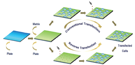
The Recent Development in Reverse Gene Transfection

Zhang Pengfei, Hu Xiufeng, Cheng Lu, Wang Wei*, Liu Wenguang*   

  1. Tianjin Key Laboratory of Composite and Functional Materials, School of Materials Science and Engineering, Tianjin University, Tianjin 300072, China
  • Received: Revised: Online: Published:
  • Supported by:

    The work was supported by the National Natural Science Foundation of China (No.51173129, 51325305).

PDF ( 932 ) Cited
Export

EndNote

Ris

BibTeX

With the in-depth study of gene therapy, the gene transfection techniques have achieved rapid development. Since it was proposed in 1997, reverse transfection concept has also aroused considerable attention. Reverse gene transfection is also known as surface-mediated transfection or matrix-mediated transfection, or solid-phase transfection. In a typical procedure of reverse transfection, DNA (or RNA) is firstly anchored to the substrate; cells are transplanted on the substrate surface in the next step; the adsorbed DNA (or RNA) is internalized into post-attached cells to realize the gene transfection. Compared with conventional gene transfection, the reverse method is imparted with several advantages including more stable complexation of vector/DNA (or RNA), more effective contact between the transfection reagent and cells, which is favourable for achieving higher transfection efficiency and lower cytotoxicity, and well-maintained transfection performance in the presence of serum. The recent research progress of reverse gene transfection and its applications are focused on in this review.

Contents
1 Introduction 2 Approaches to reverse gene transfection
2.1 Nucleic acid (and vector) directly anchored to the substrate surface
2.2 Electrostatic adsorption
2.3 Layer-by-layer
2.4 Hydrogen bond
2.5 Specific interaction
2.6 Covalent bond
3 Applications of reverse gene transfection
3.1 Tissue engineering
3.2 Gene screening
3.3 Primary cells
3.4 Non-adherent cells
4 Conclusion and perspective

CLC Number: 

[1] Palsson B, Andreadis S. Exp. Hematol., 1997, 25: 94.
[2] Ziauddin J, Sabatini D M. Nature, 2001, 411: 107.
[3] Bengali Z, Pannier A K, Segura T, Anderson B C, Jang J H, Mustoe T A, Shea L D. Biotechnol. Bioeng., 2005, 90: 290.
[4] Jewell C M, Lynn D M. Curr. Opin. Colloid Interface Sci., 2008, 13: 395.
[5] Ji Q, Yamazaki T, Hanagata N, Lee M V, Hill J P, Ariga K. Chem. Commun., 2012, 48: 8496.
[6] Wu K M, Xu J, Liu M Y, Song W, Yan J, Gao S, Zhao L Z, Zhang Y M. Int. J. Nanomed., 2013, 8: 1595.
[7] Honma K, Miyata T, Ochiya T. Curr. Drug. Discov. Tech., 2004, 1: 287.
[8] Villa-Diaz L G, Garcia-Perez J L, Krebsbach P H. Stem Cells Dev., 2010, 19: 1949.
[9] Shen H, Tan J, Saltzman W M. Nat. Mater., 2004, 3: 569.
[10] Oyane A, Wang X, Sogo Y, Ito A, Tsurushima H. Acta Biomater., 2012, 8: 2034.
[11] Nagane K, Kitada M, Wakao S, Dezawa M, Tabata Y. Tissue Eng. Part A, 2009, 15: 1655.
[12] He C X, Li N, Hu Y L, Zhu X M, Li H J, Han M, Miao P H, Hu Z J, Wang G, Liang W Q,Tabata Y, Gao J Q. Pharm. Res., 2011, 28: 1577.
[13] Solanki A, Shah S, Yin P T, Lee K B. Sci. Rep., 2013, 3: 1553.
[14] Uchimura E, Yamada S, Uebersax L, Fujita S, Miyake M, Miyake J. J. Biosci. Bioeng., 2007, 103: 101.
[15] Lin Q K, Ren K F, Ji J. Colloids Surf. B-Biointerfaces, 2009, 74: 298.
[16] Lu Z Z, Wu J, Sun T M, Ji J, Yan L F, Wang J. Biomaterials, 2008, 29: 733.
[17] Wang X F, Ren K F, Lin Q K, Ji J. Sci. China Chem., 2010, 53: 508.
[18] Yamauchi F, Kato K, Iwata H. Biochim. Biophys. Acta, 2004, 1672: 138.
[19] Holmes C A, Tabrizian M. ACS Appl. Mater. Inter., 2013, 5: 524.
[20] Tang L, Liu W, Liu G. Adv. Mater., 2010, 22: 2652.
[21] Tang L, Yang Y, Bai T, Liu W. Biomaterials, 2011, 32: 1943.
[22] Wang N, Zhang J, Sun L, Wang P, Liu W. Acta Biomater., 2014, 10: 2529.
[23] Segura T, Volk M J, Shea L D. J. Control. Release, 2003, 93: 69.
[24] Segura T, Chung P H, Shea L D. Biomaterials, 2005, 26 (13): 1575.
[25] Segura T, Shea L D. Bioconjugate Chem., 2002, 13: 621.
[26] Carbone R. Peptide Microarrays: Methods in Molecular Biology, Walker J M (Ed.).Clifton: Humana Press, 2009. 339.
[27] Park I K, Von Recum H A, Jiang S Y, Pun S H. Langmuir, 2006, 22: 8478.
[28] Zheng J, Manuel W S, Hornsby P J. Biotechnol. Progr., 2000, 16: 254.
[29] Bonadio J, Smiley E, Patil P, Goldstein S. Nat. Med., 1999, 5: 753.
[30] Yin L C, Zhao X, Ji S Z, He C. B, Wang G Y, Tang C, Gu S, Yin C. Biomaterials, 2014, 35: 2488.
[31] Elangovan S, DMello S R, Hong L, Ross R D, Allamargot C, Dawson D V, Stanford C M, Johnson G K, Sumner D R, Salem A K. Biomaterials, 2014, 35: 737.
[32] Jewell C M, Zhang J, Fredin N J, Wolff M R, Hacker T A, Lynn D M. Biomacromolecules, 2006, 7: 2483.
[33] Fujita S, Ota E, Sasaki C, Takano K, Miyake M, Miyake J. J. Biosci. Bioeng., 2007, 104: 329.
[34] Chen P C, Huang Y Y, Juang J L. Lab Chip, 2011, 11: 3619.
[35] Mannherz O, Mertens D, Hahn M, Lichter P. Genomics, 2006, 87: 665.
[36] Chia N Y, Chan Y S, Feng B, Lu X Y, Orlov Y L, Moreau D, Kumar P, Yang L, Jiang J, Lau M S, Huss M, Soh B S, Kraus P, Li P, Lufkin T, Lim B, Clarke N D, Bard F, Ng H H. Nature, 2010, 468: 316.
[37] Whitehurst A. W, Bodemann B O, Cardenas J, Ferguson D, Girard L, Peyton M, Minna J D, Hao W, Roth M G, Xie X J, White M A. Nature, 2007, 446: 815.
[38] Erfle H Neumann B, Liebel U, Rogers P, Held M, Walter T, Ellenberg J, Pepperkok R. Nat. Protoc., 2007, 2: 392.
[39] Choi S, Yu X H, Paiboonkit L J, Hollister S J, Murphy W L. Sci. Rep., 2013, 3: 1567.
[40] Kato K, Umezawa K, Miyake M, Miyake J, Nagamune T. Biotechniques, 2004, 37: 444.
[41] Li C Y, Yuan W, Jiang H, Li J S, Xu F J, Yang W T, Ma J. Bioconjugate Chem., 2011, 22: 1842.
[42] Yuan W, Li C, Zhao Y, Chen Z, Sui C G, Yang W T, Xu F J, Ma J. Adv. Fuct. Mater., 2012, 22: 1835.
[43] Bae J, Goto S, Mie M, Kobatake E. J. Biotechnol., 2010, 150: 447.
[44] Rea J C, Gibly R F, Davis N E, Barron A E, Shea L D. Biomacromolecules, 2009, 10: 2779.
[45] Uchimura E, Yamada S, Nomura T, Matsumoto K, Fujita S, Miyake M, Miyake J. J. Biosci. Bioeng., 2007, 104: 152.

[1] Xiaodong Jing, Ying Sun, Bing Yu, Youqing Shen, Hao Hu, Hailin Cong. Rational Design of Tumor Microenvironment Responsive Drug Delivery Systems [J]. Progress in Chemistry, 2021, 33(6): 926-941.
[2] Li Sidi, Hou Xin, Qi Hongzhao, Zhao Jin, Yuan Xubo. Exosomes:Provide Naturally Occurring Endogenous Nanocarriers for Effective Drug Delivery Strategies [J]. Progress in Chemistry, 2016, 28(2/3): 353-362.
[3] Liu Yajie, Zhang Peng, Du Jianwei, Wang Youxiang. Regulation the Morphology of Micro-and Nanoparticles and the Effect on Drug/Gene Delivery System [J]. Progress in Chemistry, 2016, 28(1): 67-74.
[4] Gu Xiaoxiao, Du Baoji, Li Yunhui, Gao Ying, Li Dan, Wang Erkang. Vectors Based on Nanomaterials for Gene Delivery [J]. Progress in Chemistry, 2015, 27(8): 1093-1101.
[5] Du Jianwei, Mou Yun, Wang Youxiang. Research of Ultrasound-Mediated Gene Delivery [J]. Progress in Chemistry, 2014, 26(08): 1427-1433.
[6] Yang Chanli, Dong Xiongwei, Jiang Nan, Zhang Dan, Liu Changlin*. Introduction of Metal Complex-Nucleic Acid Interactions into Cells [J]. Progress in Chemistry, 2013, 25(04): 555-562.
[7] Yang Qizhi, Liu Jia, Jiang Xulin. Application of Click Chemistry in Biomedical Polymers [J]. Progress in Chemistry, 2010, 22(12): 2377-2387.
[8] Hua Yuan Jie Ren. The Research of Functional Polymeric Gene Vector [J]. Progress in Chemistry, 2008, 20(11): 1804-1809.
[9] Hua He. Progress of the Applications of Carbon Nanotubes in Drug and Gene Delivery [J]. Progress in Chemistry, 2008, 20(11): 1798-1803.
[10]

Ji Weihang|Lin Lin|Chen Dayong|Liu Wenguang**

. Stimuli-responsive Non-viral Gene Delivery Vectors [J]. Progress in Chemistry, 2008, 20(06): 936-941.
[11] Yanqing Sun1,Jian Zhang2,Gaoyong Zhang1**,Hongxia Wang1. The Interaction between DNA and Amphoteric Surfactants [J]. Progress in Chemistry, 2006, 18(11): 1440-1445.