中文
Announcement
More
Progress in Chemistry 2015, Vol. 27 Issue (1): 91-102 DOI: 10.7536/PC140738 Previous Articles   Next Articles

• Review •

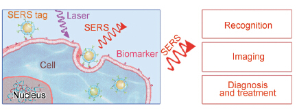
Surface-Enhanced Raman Scattering Tags Used in Cell Recognition, Imaging, Diagnosis and Treatment

Song Chunyuan*, Chen Wenqiang, Yang Yanjun, Yang Boyue, Su Shao, Wang Lianhui*   

  1. Key Lab for Organic Electronics & Information Displays (KLOEID), Institute of Advanced Materials (IAM), and Synergetic Innovation Center for Organic Electronics and Information Displays, Nanjing University of Posts & Telecommunications, Nanjing 210023, China
  • Received: Revised: Online: Published:
  • Supported by:

    The work was surpported by the National Basic Research Program of China (973 Program) (No. 2012CB933301), the National Natural Science Foundation of China (No. 61302027, 21305070, 21475064), the Natural Science Foundation of Jiangsu Province (No. BK20130871), the Program for Changjiang Scholars and Innovative Research Team in University (IRT1148), the Open Research Fund of State Key Laboratory of Bioelectronics (2013G2), Sci-tech Support Plan of Jiangsu Province (No. BE2014719), the Priority Academic Program Development of Jiangsu Higher Education Institutions (PAPD) and Foundation of Nanjing University of Posts and Telecommunications (NY212032).

PDF ( 1389 ) Cited
Export

EndNote

Ris

BibTeX

Surface-enhanced Raman scattering (SERS) refers to an abnormal surface optical phenomenon that Raman spectra of the analyte adsorbed on metal nanostructures can be significantly enhanced under laser irradiation. In recent years, SERS has been widely used in the substance detection and biological sensing, which has achieved significant development and shown great potential applications in the biomedical field. In this paper, the preparation principle and the latest development of SERS tags used in cell detection are introduced, and their recent applications in cell recognition, imaging, diagnosis and treatment are reviewed, as well as the detection strategies and issues related to SERS tag-based cell detection are discussed.

Contents
1 Introduction
2 SERS tags
2.1 Noble metal nanoparticles
2.2 Raman reporter molecules
2.3 Surface coating for protection
2.4 Biofunction with biomolecules
3 Application of SERS tags in cell
3.1 Cell recognition
3.2 Cell imaging
3.3 Cell pH sensing
3.4 Cancer diagnosis and treatment
4 Conclusions and outlooks

CLC Number: 

[1] Wu Q H. Spectrosc. Lett., 2014, 47(9): 704.
[2] Nima Z A, Mahmood M, Xu Y, Mustafa T, Watanabe F, Nedosekin D A, Juratli M A, Fahmi T, Galanzha E I, Nolan J P, Basnakian A G, Zharov V P, Biris A S.Sci. Rep., 2014, 4: 4752.
[3] Zheng X S, Hu P, Zhong J H, Zong C, Wang X, Liu B J, Ren B. Phys. Chem. C, 2014, 118(7): 3750.
[4] Fleischmann M, Hendra P J, McQuillan A J. Chem. Phys. Lett., 1974, 26(2): 163.
[5] Jeanmaire D L,Van Duyne R P. J. Electroanal. Chem. Interfacial Electrochem., 1977, 84(1): 1.
[6] Xu H, Li Q, Wang L H, He Y, Shi J Y, Tang B, Fan C H, Chem. Soc. Rev., 2014, 43: 2650.
[7] Yang X F, Li J, Pei H, Zhao Y, Zuo X L, Fan C H, Huang Q. Anal. Chem., 2014, 86(6): 3227.
[8] Zhou G B, Lin M H, Song P, Chen X Q, Chao J, Wang L H, Huang Q, Huang W, Fan C H, Zuo X L. Anal. Chem., 2014, 86(15):7843.
[9] He Y, Su Y Y, Yang X B, Kang Z H, Xu T T, Zhang R Q, Fan C H, Lee S T. J. Am. Chem. Soc., 2009, 131(12): 4434.
[10] Lu Y, Huang Q, Meng G, Wu L, Zhang J. Analyst, 2014, 139(12): 3083.
[11] Pinzaru S C, Falamas A, Dehelean C, Morari C,Venter M. Croat. Chem. Acta, 2013, 86(3): 233.
[12] Wang Y L, Li D, Li P, Wang W D, Ren W, Dong S J, Wang E K. J. Phys. Chem. C, 2007, 111(45): 16833.
[13] Otto A, Mrozek I, Grabhorn H, Akemann W J. Matter, 1992, 4: 1143.
[14] Wu D Y, Liu X M, Duan S, Xu X, Ren B, Lin S H, Tian Z Q. Phys. Chem., 2008, 112(11): 4195.
[15] Xu H, Aizpurua J, Käll M, Apell P. Phys. Rev. E, 2000, 62(3): 4318.
[16] Fang W, Wang Z Y, Zong S F, Chen H, Zhu D, Zhong Y, Cui Y P. Biosens. Bioelectron., 2014, 57: 10.
[17] DeVetter B M, Bhargava R, Murphy C J. Photochem. Photobiol., 2014, 90(2): 415.
[18] Zhu H, Du M L, Zhang M, Wang P, Bao S Y, Zou M L, Fu Y Q, Yao J M. Biosens. Bioelectron., 2014, 54: 91.
[19] Wang Y, Li D, Li P, Wang W, Ren W, Dong S, Wang E. J. Phys. Chem. C, 2007, 111(45): 16833.
[20] Wang Z Y, Zong S F, Yang J, Song C Y, Li J, Cui Y P. Biosens. Bioelectron., 2010, 26(1): 241.
[21] Jana N R, Gearheart L, Murphy C J. J. Phys. Chem. B, 2001, 105(19): 4065.
[22] Wang C G, Irudayaraj J. Small, 2010, 6(2): 283.
[23] Yuan H, Khoury C G, Hwang H, Wilson C M, Grant G A, Vo-Dinh T. Nanotechnol., 2012, 23(7): 075102.
[24] Boca S, Rugina D, Pintea A, Barbu-Tudoran L, Astilean S. Nanotechnol., 2011, 22(5): 055702.
[25] Pavan Kumar G V, Shruthi S, Vibha B. J. Phys. Chem. C, 2007, 111(11): 4388.
[26] Lee S, Chon H, Lee M, Choo J, Shin S Y, Lee Y H, Rhyu I J, Son S W, Oh C H. Biosens.Bioelectron., 2009, 24(7): 2260.
[27] Song C, Wang Z, Yang J. Acta Chim. Sinica, 2009, 67: 493.
[28] Wang X M, Zhang R Y, Wu C H, Dai Y Y, Song M, Gao F, Lv G, Li J Y, Li X M. J. Biomed. Mater. Res. Part A, 2007, 80(4): 852.
[29] Tan X B, Wang Z Y, Yang J, Song C Y, Zhang R H, Cui Y P. Nanotechnol., 2009, 20(44): 445102.
[30] Wang Z Y, Wu H, Wang C L, Xu S H, Cui Y P. J. Mater. Chem., 2011, 21(12): 4307.
[31] Fang H, Zhang C X, Liu L, Zhao Y M, Xu H J, Biosens. Bioelectron., 2014, 64: 434.
[32] Chen Z Y, Dai Z M, Chen N, Liu S P, Pang F F, Lu B, Wang T Y. IEEE Photonics Technol. Lett, 2014, 26(8): 777.
[33] Deng Y L, Juang Y J. Biosens. Bioelectron., 2014, 53: 37.
[34] Sarkar A, Wang H, Daniels-Race T. Electron. Mater. Lett., 2014, 10(2): 325.
[35] MacLaughlin C M, Mullaithilaga N, Yang G, Ip S Y, Wang C, Walker G C. Langmuir, 2013, 29(6): 1908.
[36] Guven B, Dudak F C, Boyaci I H, Tamer U, Ozsoz M. Analyst, 2014, 139(5): 1141.
[37] Lin C C, Chang C W. Biosens. Bioelectron., 2014, 51: 297.
[38] You L J, An Q, Guo J, Hu J J, Wang C C. RSC Adv., 2013, 3(38): 17469.
[39] Furusho H, Oishi M, Kishi T, Yasumori A, Nagasaki Y. Chem. Lett., 2010, 39(1): 52.
[40] Kang H, Yim J, Jeong S, Yang J K, Kyeong S, Jeon S J, Kim J, Eom K D, Lee H, Kim H I, Jeong D H, Kim J H, Lee Y S. ACS Appl. Mater. Interfaces, 2013, 5(24): 12804.
[41] Wrzesien J, Graham D. Tetrahedron, 2012, 68(4): 1230.
[42] Brady C I, Mack N H, Brown L O, Doorn S K. Anal. Chem. Phys. Lett., 2009, 81: 7181.
[43] Mulvaney S P, Musick M D, Keating C D, Natan M J. Langmuir, 2003, 19: 4784.
[44] Pinkhasova P, Yang L, Zhang Y, Sukhishvili S, Du H. Langmuir, 2012, 28: 2529.
[45] Niu X J, Chen H Y, Wang Y Q, Wang W H, Sun X Y, Chen L X. ACS Appl. Mater. Interfaces, 2014, 6(7): 5152.
[46] Zhang D M, Ansar S M, Vangala K, Jiang D P. J. Raman Spectrosc., 2010, 41(9): 952.
[47] Dey P, Olds W, Blakey I, Thurecht K J, Izake E L, Fredericks P M. J. Raman Spectrosc., 2013, 44(12): 1659.
[48] Sun L, Sung K B, Dentinger C, Lutz B, Nguyen L. B. Nano Lett., 2007, 7: 351.
[49] Dinish U S, Balasundaram G, Chang Y T, Olivo M. Sci. Rep., 2014, 4: 4075.
[50] Dinish U S, Fu C Y, Soh K S, Bhuvaneswari R, Kumar A, Olivo M. Biosens. Bioelectron., 2012, 33(1): 293.
[51] Wang X, Qian X M, Beitler J J, Chen Z G, Khuri F R, Lewis M M, Shin H J C, Nie S M, Shin D M. Cancer Res., 2011, 71(5): 1526.
[52] Liu Z M, Guo Z Y, Zhong H Q, Qin X C, Wan M M, Yang B W. Phys. Chem. Chem. Phys., 2013, 15(8): 2961.
[53] Marz A, Trupp S, Rosch P, Mohr G J, Popp J. Anal. Bioanal.Chem., 2012, 402(8): 2625.
[54] MacLaughlin C M, Parker E P K, Walker G C, Wang C. Nanomed. Nanotechnol. Biol. Med., 2013, 9(1): 55.
[55] Sha M Y, Xu H X, Natan M J, Cromer R. J. Am. Chem. Soc., 2008, 130(51): 17214.
[56] Zong S F, Wang Z Y, Yang J, Wang C L, Xu S H, Cui Y P. Talanta, 2012, 97: 368.
[57] Samanta A, Jana S, Das R K, Chang Y T. RSC Adv., 2014, 4(24): 12415.
[58] Chen H, Wang Z Y, Ma X Q, Zong S F, Cui Y P. Talanta, 2013, 116: 978.
[59] Vendrell M, Maiti K K, Dhaliwal K, Chang Y T. Trends Biotechnol., 2013, 31(4): 249.
[60] Lee S. Anal. Chem., 2007, 79: 916.
[61] Lee S, Chon H, Lee M, Choo J, Shin S Y, Lee Y H, Rhyu I J, Son S W, Oh C H. Biosens. Bioelectron., 2009, 24(7): 2260.
[62] Wu P, Gao Y, Zhang H, Cai C X. Anal. Chem., 2012, 84(18): 7692.
[63] Zhang G, Qu G, Chen Y, Shen A, Xie W, Zhou X, Hu J. J. Phys. Chem. B, 2013, 1(35): 4364.
[64] Wu P, Gao Y, Lu Y, Zhang H, Cai C. Analyst, 2013, 138(21): 6501.
[65] Zhang Y Y, Yu W S, Pei L, Lai K Q, Rasco B A, Huang Y Q. Food Chem., 2014, 169: 80.
[66] Ganbold E O, Lee C M, Cho E M, Son S J, Kim S, Joo S W, Yang S I. Anal. Methods, 2014, 6(11): 3573.
[67] Zong S, Wang Z, Yang J, Wang C, Xu S, Cui Y. Talanta, 2012, 97: 368.
[68] 崔颜 (Cui Y), 任斌 (Ren B), 田中群 (Tian Z Q). 东南大学学报 (医学版) (Journal of Southeast University (Medical Science Edition)), 2011, 1: 254.
[69] Kneipp J, Kneipp H, Wittig B. J. Phys. Chem. C, 2010, 114: 7421.
[70] Zong S, Wang Z, Yang J, Cui Y. Anal. Chem., 2011, 83(11): 4178.
[71] Liang L, Li J, Li Q, Huang Q, Shi J Y, Yan H, Fan C H. Angew. Chem. Int. Edit., 2014, 53(30): 7745
[72] Song J, Zhou J, Duan H. J. Am. Chem. Soc., 2012, 134(32): 13458.
[73] Wang Y Q, Chen L X, Liu P. Chem. Eur. J., 2012, 18(19): 5935.
[74] Lu W, Singh A K, Khan S A. J. Am. Chem. Soc., 2010,132(51): 18103.
[75] Shen S, Tang H Y, Zhang X T, Ren J F, Pang Z Q, Wang D G, Gao H L, Qian Y, Jiang X G, Yang W L. Biomater., 2013, 34(12): 3150.
[76] Fu C C, Gu Y J, Wu Z Y, Wang Y Y, Xu S P, Xu W Q. Sens. Actuators B, 2014, 201: 173.

[1] Jing He, Jia Chen, Hongdeng Qiu. Synthesis of Traditional Chinese Medicines-Derived Carbon Dots for Bioimaging and Therapeutics [J]. Progress in Chemistry, 2023, 35(5): 655-682.
[2] Zixuan Liao, Yuhui Wang, Jianping Zheng. Research Advance of Carbon-Dots Based Hydrophilic Room Temperature Phosphorescent Composites [J]. Progress in Chemistry, 2023, 35(2): 263-373.
[3] Anchen Fu, Yanjia Mao, Hongbo Wang, Zhijuan Cao. Development and Application of Dioxetane-based Chemiluminescent Probes [J]. Progress in Chemistry, 2023, 35(2): 189-205.
[4] Dang Zhang, Xi Wang, Lei Wang. Biomedical Applications of Enzyme-Powered Micro/Nanomotors [J]. Progress in Chemistry, 2022, 34(9): 2035-2050.
[5] Feng Lu, Ting Zhao, Xiaojun Sun, Quli Fan, Wei Huang. Design of NIR-Ⅱ Emissive Rare-earth Nanoparticles and Their Applications for Bio-imaging [J]. Progress in Chemistry, 2022, 34(6): 1348-1358.
[6] Zhen Wang, Xi Li, Yuanyuan Li, Qi Wang, Xiaomei Lu, Quli Fan. Activatable NIR-Ⅱ Probe for Tumor Imaging [J]. Progress in Chemistry, 2022, 34(1): 198-206.
[7] Xuechuan Wang, Yansong Wang, Qingxin Han, Xiaolong Sun. Small-Molecular Organic Fluorescent Probes for Formaldehyde Recognition and Applications [J]. Progress in Chemistry, 2021, 33(9): 1496-1510.
[8] Huifeng Xu, Yongqiang Dong, Xi Zhu, Lishuang Yu. Novel Two-Dimensional MXene for Biomedical Applications [J]. Progress in Chemistry, 2021, 33(5): 752-766.
[9] Yecheng Dang, Yangzhen Feng, Dugang Chen. Red/Near-Infrared Biothiol Fluorescent Probes [J]. Progress in Chemistry, 2021, 33(5): 868-882.
[10] Chao Zhao, Zongwei Cai. Mass Spectrometry Imaging and Omics for Environmental Toxicology Research [J]. Progress in Chemistry, 2021, 33(4): 503-511.
[11] Yunxue Wu, Hengyi Zhang, Yu Liu. Application of Azobenzene Derivative Probes in Hypoxia Cell Imaging [J]. Progress in Chemistry, 2021, 33(3): 331-340.
[12] Pingping Zhao, Junxing Yang, Jianhui Shi, Jingyi Zhu. Construction and Application of Dendrimer-Based SPECT Imaging Agent [J]. Progress in Chemistry, 2021, 33(3): 394-405.
[13] Fei Ren, Jianbing Shi, Bin Tong, Zhengxu Cai, Yuping Dong. Near Infrared Fluorescent Dyes with Aggregation-Induced Emission [J]. Progress in Chemistry, 2021, 33(3): 341-354.
[14] Yafang Sun, Ziping Zhou, Tong Shu, Lisheng Qian, Lei Su, Xueji Zhang. Multicolor Luminescent Gold Nanoclusters: From Structure to Biosensing and Bioimaging [J]. Progress in Chemistry, 2021, 33(2): 179-187.
[15] Yuanyuan Liu, Yun Guo, Xiaogang Luo, Genyan Liu, Qi Sun. Detection of Metal Ions, Small Molecules and Large Molecules by Near-Infrared Fluorescent Probes [J]. Progress in Chemistry, 2021, 33(2): 199-215.