 PDF(18489 KB)
 PDF(18489 KB)
PDF(18489 KB)
PDF(18489 KB)
基于碳点的发光材料在潜在手印显现中的应用
 ({{custom_author.role_cn}}), {{javascript:window.custom_author_cn_index++;}}
Application of Luminescent Materials Based on Carbon Dots in Development of Latent Fingerprints
({{custom_author.role_en}}), {{javascript:window.custom_author_en_index++;}}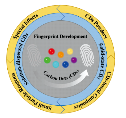
| {{custom_ref.label}} |
{{custom_citation.content}}
{{custom_citation.annotation}}
|
/
| 〈 |
|
〉 |


 AI Summary

