 PDF(29530 KB)
 PDF(29530 KB)
PDF(29530 KB)
PDF(29530 KB)
中药碳点的合成及其在生物成像和医学治疗方面的应用
 ({{custom_author.role_cn}}), {{javascript:window.custom_author_cn_index++;}}
Synthesis of Traditional Chinese Medicines-Derived Carbon Dots for Bioimaging and Therapeutics
({{custom_author.role_en}}), {{javascript:window.custom_author_en_index++;}}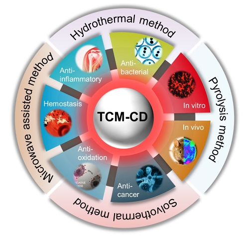
| {{custom_ref.label}} |
{{custom_citation.content}}
{{custom_citation.annotation}}
|
/
| 〈 |
|
〉 |


 AI Summary

