中文
Announcement
More
Progress in Chemistry 2021, Vol. 33 Issue (3): 341-354 DOI: 10.7536/PC200614 Previous Articles   Next Articles

• Review •

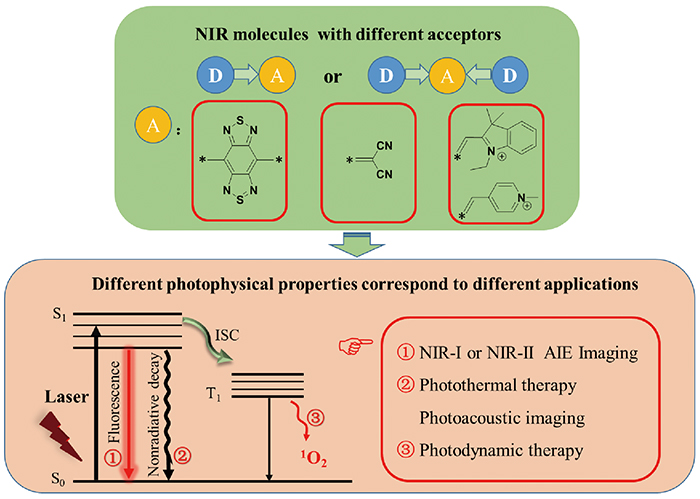
Near Infrared Fluorescent Dyes with Aggregation-Induced Emission

Fei Ren1, Jianbing Shi1,*(), Bin Tong1, Zhengxu Cai1, Yuping Dong1,*()   

  1. 1 School of Materials Science and Engineering, Beijing Institute of Technology,Beijing 100081, China
  • Received: Revised: Online: Published:
  • Contact: Jianbing Shi, Yuping Dong
  • Supported by:
    the National Natural Science Foundation of China(21875019); the National Natural Science Foundation of China(51673024); the National Natural Science Foundation of China(21975020); the National Natural Science Foundation of China(51803009)
Richhtml ( 59 ) PDF ( 1157 ) Cited
Export

EndNote

Ris

BibTeX

The discovery of the aggregation-induced emission(AIE) phenomenon provides the best solution to solve the problem of fluorescence quenching of traditional organic fluorescent molecules at high concentrations and aggregation state. AIE molecules are widely used in many fields such as photoelectric devices, chemical sensing, biological imaging and targeting therapy. With the deepening of the research on the emissive mechanism of AIE, the AIE molecular system has been greatly expanded. Among them, a class of AIE molecules with donor-acceptor structures can significantly reduce the molecular energy gap and extend the emission wavelengths of molecules from the visible light region(400~700 nm) to the near infrared(NIR) region(700~1700 nm). Due to the unique advantages of NIR fluorescent molecules in the field of biomedicine, they have become the hot topic of AIE research. With the continuous exploration of the design and application of NIR molecules, AIE molecules with different functions and longer emission wavelengths have also been developed, and realized the application of NIR fluorescence imaging, photoacoustic imaging, photodynamic therapy and photothermal therapy to specific tissues of organisms. This article summarizes the structure of NIR fluorescent molecules with AIE performance in recent years and their related applications in the field of biomedicine.

Contents

1 Introduction

2 The discovery and mechanism of aggregation-induced emission

3 The advantages of NIR and the partition of fluorescent windows

4 Principle of molecular design of NIR dyes

5 NIR fluorescent dyes with AIE property and their applications

5.1 Design and application of benzothiadiazole NIR dyes

5.2 Design and application of malononitrile NIR dyes

5.3 Design and application of ionic NIR dyes

6 Conclusion

Fig.1 (a) Tissue penetration depth of light with different wavelengths;(b) light when entering a tissue can be reflected or absorbed by molecules within the tissue or excite fluorophores to emit light at different wavelengths[12]. Copyright 2018, Royal Society of Chemistry
Fig.2 (a) Chemical structure of TB1;(b) NIR-Ⅱ fluorescence images of TB1 and IR-26 in solution and powder states(solution: DMSO);(c) UV absorption and fluorescence spectra of TB1 in THF solution;(d) fluorescence intensity ratio of TB1 in THF/water mixture with different water contents;(e) preparation route of TB1-RGD nanoparticle;(f) brain non-invasive NIR-Ⅱ fluorescence imaging on mice with intact scalp and skull(808 nm, 60 mW·cm-2)[35]. Copyright 2018, Wiley
Fig.3 (a) Synthesis route of HLZ-BTED;(b) fluorescence emission spectra of HLZ-BTED at different water contents(0% to 95%)(Ex: 785 nm);(c) the NIR-Ⅱ tumor blood vessel fluorescence images of 4T1 tumor-bearing mice 2 minutes after injection of HLZ-BTED nanoparticles into the tail vein[36]. Copyright 2019, Royal Society of Chemistry
Fig.4 (a) The chemical structure of HQL1 and HQL2;(b) UV-vis-NIR absorption spectra and NIR-Ⅱ fluorescence spectra of HQL1 and HQL2 in DCM;(c) fluorescence imaging of U87MG tumor-bearing nude mice(808 nm, 90 mW·cm-2);(d) cross-sectional intensity of capillaries imaged by HQL2 nanoparticles(black line) and Gaussian fitted fluorescence intensity distribution map(red line)(red dotted line in c)[37].Copyright 2020, Royal Society of Chemistry
Fig.5 (a) The chemical structure of the target molecule;(b~d) NIR-Ⅱ and NIR-Ⅱb fluorescence images of cerebral vasculature with different LP filters in C57BL/6 mice(n = 3) after tail intravenous injection of HL3 dots(200 μL, 1.5 mg·mL -1):(b) 1000 nm LP, 4 ms exposure time, and 90 mW·cm-2;(c) 1250 nm LP, 60 ms exposure time, and 90 mW·cm-2;(d) 1550 nm LP, 500 ms exposure time, and 90 mW·cm-2;(e~g) the fluorescence intensity profiles fitted using Gaussian, cross-section intensity(black lines), and the tiny vessel(red-dashed lines)[38]. Copyright 2020, Royal Society of Chemistry
Fig.6 (a) The chemical structure of the target molecule 2TT-oC6B and the schematic diagram of the preparation of AIE nanoparticles;(b) UV absorption and fluorescence emission spectra of 2TT-oC6B nanoparticles in deionized water;(c) intraureteral injection of 2TT-oC6B nanoparticles to achieve NIR-Ⅱ fluorescence imaging of the ureter in the rabbit model(Ex: 808 nm);(d) comparison of 2TT-oC6B nanoparticles(red dotted line) and ICG(blue dotted line) on ureter imaging quality[39]. Copyright 2020, American Chemical Society
Fig.7 (a) Chemical structure of SYL molecule and NIR-Ⅱ fluorescence pictures in visible light and ultraviolet light;(b) after injection of SYL nanoparticles into the tail vein of 4T1 tumor-bearing mice for 0, 2, 8, 12 and 24 h, in vivo NIR-Ⅱ fluorescence and PA images(808 nm, 82 mW·cm-2)[40]. Copyright 2019, Royal Society of Chemistry
Fig.8 Chemical structure of photosensitizer TQ-BTPE and schematic illustration of NIR-Ⅱ light activated two-photon photodynamic cancer cell ablation[41]. Copyright 2020, Wiley
Fig.9 (a) Target molecular structures and single crystal structures of QM-1, QM-2, and QM-3;(b) in vivo non-invasive imaging of tumor-bearing mice at different time after intravenous injection of QM-5(0.15 mg/kg);(c) 3D fluorescence imaging of tumor-bearing mice after the tail vein injection of QM-5(0.15 mg/kg) for 24 hours[48]. Copyright 2015, Wiley
Fig.10 Rational design of NIR AIE-active probes for Aβ deposition. (a) Commercial probe ThT based on thealways-on pattern;(b,c) the“step-by-step” strategy to address the inherent defects of commercial ThT and create ultrasensitive off-on NIR probes:(i) introducing lipophilic π-conjugated thiophene-bridge for extending the wavelength to the NIR region with BBB penetrability,(ii) replacing the ACQ to AIE building block, and(iii) tuning the sulfonate substituted position for guaranteeing fluorescence-off state before binding to Aβ deposition.;(d~g) histological staining of the brain slices in the hippocampus region from wild-type mice and Alzheimer’s disease(AD)-model(APP/PS1 transgenic) mice using ThS and QM-FN-SO3, respectively;(h) the intensity profiles of the linear regions of interest(ROI) crossing the brain slices;(i) the S/N ratios of DCM-N, QM-FN, and QM-FN-SO3[49]. Copyright 2019, American Chemical Society
Fig.11 (a) Chemical synthesis routes of TFM;(b) preparation of TFM nanoparticles by nanodeposition method;(c) illustration of the use of TFM NPs for PAI-guided PTT-PDT cancer theranostics[50]. Copyright 2019, Wiley
Fig.12 (a) Synthesis routes of the target molecules;(b) schematic diagram of the NIR afterglow luminescence mechanism of AGL AIE dots;(c) schematic diagram of cancer surgery guided by NIR afterglow imaging[51]. Copyright 2019, American Chemical Society
Fig.13 Design and characterization of the developed TPACN-D-Ala probe for bacterial tracking and photodynamic therapy(PDT);(a) TPACN-D-Ala was synthesized by combining D-Ala with AIE photosensitizer TPACN, which could be intravenously injected into the bacteria-infected mouse to realize specific fluorescence light-up and image-guided antibacterial PDT;(b) Once reaching the infected tissue site through blood circulation, TPACN-D-Ala would be integrated into peptidoglycan to produce intense NIR fluorescence, specific imaging and efficient treatment of invasive bacteria could be achieved in vivo;(c) UV absorption and fluorescence emission of TPACN-D-Ala or TPACN in PBS;(d) measurement of 1O2 production of TPACN-D-Ala(10 μM) or Ce6(10 μM) using ADBA under light irradiation(white light, 60 mW·cm -2). ABDA(black) solutions were used as control; A0 and A are the absorbance of ABDA at 378 nm[52]. Copyright 2020, Royal Society of Chemistry
Fig.14 Preparation of UCNP@TTD cRGD NPs; schematic diagram of NIR fluorescence imaging and PDT application of deep tumors in 3D cancer cell spheres and mouse tumor models in vitro under NIR laser irradiation[53]. Copyright 2019, Ivyspring
Fig.15 (a) The chemical structures of the target molecules;(b) real-time video screenshot of TPP-1, TPP-2 and TPP-3 co-cultured with HeLa under the same conditions [TPP-1] = [TPP-2] = [TPP-3] = 1.0×10⁻⁷ mol/L[54]. Copyright 2018, Royal Society of Chemistry
Fig.16 (a) Schematic diagram of PMTi synthesis;(b) mitochondrial-targeted subcellular drug delivery[55]. Copyright 2019, Wiley
Fig.17 (a) The chemical structure of TPE-DPA-TCyP;(b) the proposed mechanism of TPE-DPA-TCyP as an effective ICD inducer for antitumor immunity[56]. Copyright 2019, Wiley
Fig.18 (a) Chemical structures of target molecules;(b) relative emission intensity(I/I0) spectra of different solvent ratios, inset: fluorescence photos of TTPy in DMSO solution and DMSO/toluene mixture(containing 95% toluene) under UV irradiation at 365 nm;(c) the volume growth curves of tumors at different time points post-treatment in different groups;(d) body weight measurement of the mice in each group[57]. Copyright 2018, Wiley
Fig.19 (a) Chemical structures of TFPy, TFVP and TPE-TFPy;(b) schematic illustration of using three AIEgens for achieving “1+1+1>3” synergistic enhanced photodynamic therapy [58]. Copyright 2020, Wiley
Fig.20 (a) The chemical structure of the target molecule; overlaid confocal images of AS2CP-TPA-stained HeLa cells before(pseudo red) and after(pseudo green) incubation in PBS containing 100 μM Hg 2+ for 40 min(b, c) or incubation in PBS only for 40 min(d, e)(λex = 488 nm and λ em= 600~750 nm)[59]. Copyright 2018, Royal Society of Chemistry
Fig.21 Schematic illustration of using AIEgen TTVP for ultrafast discrimination of Gram-positive bacteria and highly efficient photodynamic antibacterial therapy[60]. Copyright 2020, Elsevier
[1]
Luo J D, Xie Z L, Lam J W Y, Cheng L, Tang B Z, Chen H Y, Qiu C F, Kwok H S, Zhan X W, Liu Y Q, Zhu D B. Chem. Commun., 2001, (18):1740.
[2]
Fan X, Sun J L, Wang F Z, Chu Z Z, Wang P, Dong Y Q, Hu R R, Tang B Z, Zou D C. Chem. Commun., 2008, (26):2989.
[3]
Feng H T, Yuan Y X, Xiong J B, Zheng Y S, Tang B Z. Chem. Soc. Rev., 2018, 47(19):7452.
[4]
Zhang Y H, Mao H L, Xu W Q, Shi J B, Cai Z X, Tong B, Dong Y P. Chem. Eur. J., 2018, 24(60):15965.
[5]
Feng X, Tong B, Shen J B, Shi J B, Han T, Chen L, Zhi J G, Lu P, Ma Y G, Dong Y P. J. Phys. Chem. B, 2010, 114(50):16731.
[6]
He J T, Xu B, Chen F P, Xia H J, Li K P, Ye L, Tian W J. J. Phys. Chem. C, 2009, 113(22):9892.
[7]
Mei J, Hong Y N, Lam J W Y, Qin A J, Tang Y H, Tang B Z. Adv. Mater., 2014, 26(31):5429.
[8]
Hu R R, Leung N L C, Tang B Z. Chem. Soc. Rev., 2014, 43(13):4494.
[9]
Wang D, Tang B Z. Acc. Chem. Res., 2019, 52:2559.
[10]
Guo Z Q, Park S, Yoon J, Shin I. Chem. Soc. Rev., 2014, 43(1):16.
[11]
Gao M, Yu F B, Lv C, Choo J, Chen L X. Chem. Soc. Rev., 2017, 46(8):2237.
[12]
He S Q, Song J, Qu J L, Cheng Z. Chem. Soc. Rev., 2018, 47(12):4258.
[13]
Hong G S, Antaris A L, Dai H J. Nat. Biomed. Eng., 2017, 1:0010.
[14]
Wang X D, Wang S M, Lv J, Shao S Y, Wang L X, Jing X B, Wang F S. Chem. Sci., 2019, 10(10):2915.
[15]
Huang G X, Xia Q, Huang W B, Tian J W, He Z K, Li B S, Tang B Z. Angew. Chem. Int. Ed., 2019, 58(49):17814.
[16]
Guo X M, Yuan P S, Qiao X F, Yang D Z, Dai Y F, Sun Q, Qin A J, Tang B Z, Ma D G. Adv. Funct. Mater., 2020, 30(9):1908704.
[17]
Wei G Q, Tao Y C, Wu J J, Li Z Z, Zhuo M P, Wang X D, Liao L S. J. Phys. Chem. Lett., 2019, 10:679.
[18]
Shahbazi M A, Faghfouri L, Ferreira M P A, Figueiredo P, Maleki H, Sefat F, Hirvonen J, Santos H A. Chem. Soc. Rev., 2020, 49(4):1253.
[19]
Cheng H B, Li Y Y, Tang B Z, Yoon J. Chem. Soc. Rev., 2020, 49(1):21.
[20]
Mei J, Leung N L C, Kwok R T K, Lam J W Y, Tang B Z. Chem. Rev., 2015, 115(21):11718.
[21]
Hong Y N, Lam J W Y, Tang B Z. Chem. Commun., 2009(29):4332.
[22]
Hong Y N, Lam J W Y, Tang B Z. Chem. Soc. Rev., 2011, 40(11):5361.
[23]
Ding D, Li K, Liu B, Tang B Z. Acc. Chem. Res., 2013, 46(11):2441.
[24]
Chi Z G, Zhang X Q, Xu B J, Zhou X, Ma C P, Zhang Y, Liu S W, Xu J R. Chem. Soc. Rev., 2012, 41(10):3878.
[25]
Tu Y J, Liu J K, Zhang H K, Peng Q, Lam J W Y, Tang B Z. Angew. Chem. Int. Ed., 2019, 58(42):14911.
[26]
Tian R, Ma H, Yang Q, Wan H, Zhu S, Chandra S, Sun H, Kiesewetter D O, Niu G, Liang Y, Chen X. Chem. Sci. 2019, 10:326.
[27]
Zhang X, An L, Tian Q W, Lin J M, Yang S P. J. Mater. Chem. B, 2020, 8(22):4738.
[28]
Staderini M, Martín M A, Bolognesi M L, MenÉndez J C. Chem. Soc. Rev., 2015, 44(7):1807.
[29]
Kaur M, Choi D H. Chem. Soc. Rev., 2015, 44(1):58.
[30]
Sun W, Guo S G, Hu C, Fan J L, Peng X J. Chem. Rev., 2016, 116(14):7768.
[31]
Kowada T, Maeda H, Kikuchi K. Chem. Soc. Rev., 2015, 44(14):4953.
[32]
Toutchkine A, Nguyen D V, Hahn K M. Org. Lett., 2007, 9(15):2775.
[33]
Samanta A, Vendrell M, Das R, Chang Y T. Chem. Commun., 2010, 46(39):7406.
[34]
Ding F, Zhan Y B, Lu X J, Sun Y. Chem. Sci., 2018, 9(19):4370.
[35]
Sheng Z H, Guo B, Hu D H, Xu S D, Wu W B, Liew W H, Yao K, Jiang J Y, Liu C B, Zheng H R, Liu B. Adv. Mater., 2018, 30(29):1870214.
[36]
Lin J C, Zeng X D, Xiao Y L, Tang L, Nong J X, Liu Y F, Zhou H, Ding B B, Xu F C, Tong H X, Deng Z X, Hong X C. Chem. Sci., 2019, 10(4):1219.
[37]
Li Q Q, Ding Q H, Li Y, Zeng X D, Liu Y S, Lu S Y, Zhou H, Wang X F, Wu J Z, Meng X L, Deng Z X, Xiao Y L. Chem. Commun., 2020, 56(22):3289.
[38]
Li Y, Liu Y F, Li Q Q, Zeng X D, Tian T, Zhou W Y, Cui Y, Wang X K, Cheng X D, Ding Q H, Wang X F, Wu J Z, Deng H, Li Y Q, Meng X L, Deng Z X, Hong X C, Xiao Y L. Chem. Sci., 2020, 11(10):2621.
[39]
Du J, Liu S J, Zhang P F, Liu H X, Li Y Y, He W, Li C B, Chau J H C, Kwok R T K, Lam J W Y, Cai L T, Huang Y H, Zhang W J, Hou J Q, Tang B Z. ACS Appl. Mater. Interfaces, 2020, 12(7):8040.
[40]
Zhang R P, Xu Y L, Zhang Y, Kim H S, Sharma A, Gao J, Yang G F, Kim J S, Sun Y. Chem. Sci., 2019, 10(36):8348.
[41]
Wang S W, Chen H, Liu J, Chen C J, Liu B. Adv. Funct. Mater., 2020, 30(30):2002546.
[42]
Redon S, Eucat G, Ipuy M, Jeanneau E, Gautier-Luneau I, Ibanez A, Andraud C, Bretonnière Y. Dye. Pigment., 2018, 156:116.
[43]
Ren F, Shi J B, Tong B, Cai Z X, Dong Y P. Mater. Chem. Front., 2019, 3(10):2072.
[44]
Li X J, Yao J, Angunawela I, Sun C K, Xue L W, Liebman-Pelaez A, Zhu C H, Yang C H, Zhang Z G, Ade H, Li Y F. Adv. Energy Mater., 2018, 8(23):1800815.
[45]
Yang Y K, Qiu B B, Chen S S, Zhou Q J, Peng Y, Zhang Z G, Yao J, Luo Z H, Chen X F, Xue L W, Feng L L, Yang C, Li Y F. J. Mater. Chem. A, 2018, 6(20):9613.
[46]
Guo Z Q, Shao A D, Zhu W H. J. Mater. Chem. C, 2016, 4(14):2640.
[47]
Lei Y X, Lai Y Y, Dong L C, Shang G J, Cai Z X, Shi J B, Zhi J G, Li P F, Huang X B, Tong B, Dong Y P. Chem. Eur. J., 2018, 24(2):434.
[48]
Shao A D, Xie Y S, Zhu S J, Guo Z Q, Zhu S Q, Guo J, Shi P, James T D, Tian H, Zhu W H. Angew. Chem. Int. Ed., 2015, 54(25):7275.
[49]
Fu W, Yan C, Guo Z, Zhang J, Zhang H, Tian H, Zhu W H. J. Am. Chem. Soc., 2019, 141:3171.
[50]
Wang D, Lee M M S, Xu W H, Shan G G, Zheng X Y, Kwok R T K, Lam J W Y, Hu X L, Tang B Z. Angew. Chem., 2019, 131(17):5684.
[51]
Ni X, Zhang X Y, Duan X C, Zheng H L, Xue X S, Ding D. Nano Lett., 2019, 19(1):318.
[52]
Mao D, Hu F, Kenry, Qi G B, Ji S L, Wu W B, Kong D L, Liu B. Mater. Horiz., 2020, 7(4):1138.
[53]
Jin G R, He R Y, Liu Q, Lin M, Dong Y Q, Li K, Tang B Z, Liu B, Xu F. Theranostics, 2019, 9(1):246.
[54]
Ren F, Liu P, Gao Y, Shi J B, Tong B, Cai Z X, Dong Y P. Mater. Chem. Front., 2019, 3(1):57.
[55]
Chen Y Z, Ai W T, Guo X, Li Y W, Ma Y F, Chen L F, Zhang H, Wang T X, Zhang X, Wang Z. Small, 2019, 15(30):1902352.
[56]
Chen C, Ni X, Jia S R, Liang Y, Wu X L, Kong D L, Ding D. Adv. Mater., 2019, 31(52):1970372.
[57]
Wang D, Lee M M S, Shan G G, Kwok R T K, Lam J W Y, Su H F, Cai Y C, Tang B Z. Adv. Mater., 2018, 30(39):1802105.
[58]
Xu W H, Lee M M S, Nie J J, Zhang Z H, Kwok R T K, Lam J W Y, Xu F J, Wang D, Tang B Z. Angew. Chem. Int. Ed., 2020, 59(24):9610.
[59]
Zhang W J, Yu C Y Y, Kwok R T K, Lam J W Y, Tang B Z. J. Mater. Chem. B, 2018, 6(10):1501.
[60]
Lee M M S, Xu W H, Zheng L, Yu B R, Leung A C S, Kwok R T K, Lam J W Y, Xu F J, Wang D, Tang B Z. Biomaterials, 2020, 230:119582.
[1] Yaoyao Li, Jingmin Liu, Guozhen Fang, Dongdong Zhang, Qinghua Wang, Shuo Wang. Biosensor Detection and Imaging Based on Persistence Luminescence Nanoprobe [J]. Progress in Chemistry, 2017, 29(6): 667-682.