 PDF(12463 KB)
 PDF(12463 KB)
PDF(12463 KB)
PDF(12463 KB)
基于生物素的荧光有机小分子及其应用
 ({{custom_author.role_cn}}), {{javascript:window.custom_author_cn_index++;}}
Fluorescent Organic Small Molecule Based on Biotin and Their Applications
({{custom_author.role_en}}), {{javascript:window.custom_author_en_index++;}}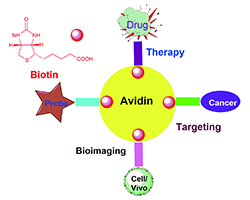
| {{custom_ref.label}} |
{{custom_citation.content}}
{{custom_citation.annotation}}
|
/
| 〈 |
|
〉 |


 AI Summary

