English
新闻公告
More
化学进展 2024, Vol. 36 Issue (2): 271-284 DOI: 10.7536/PC230610 前一篇   

• 综述 •

离子掺杂介孔生物活性玻璃的合成及应用研究

李其伟, 廖建国*()   

  1. 河南理工大学材料科学与工程学院 焦作 454000
  • 收稿日期:2023-06-10 修回日期:2023-10-25 出版日期:2024-02-24 发布日期:2024-01-08
  • 基金资助:
    河南省科技攻关项目(222102310112); 河南省科技攻关项目(222102320028)

Synthesis and Application of Ion-Doped Mesoporous Bioactive Glasses

Qiwei Li, Jianguo Liao()   

  1. School of Materials Science and Engineering, Henan Polytechnic University, Jiaozuo 454000, China
  • Received:2023-06-10 Revised:2023-10-25 Online:2024-02-24 Published:2024-01-08
  • Contact: *e-mail: liaojianguo10@hpu.edu.cn
  • Supported by:
    Science and Technology Research Project of Henan Province(222102310112); Science and Technology Research Project of Henan Province(222102320028)

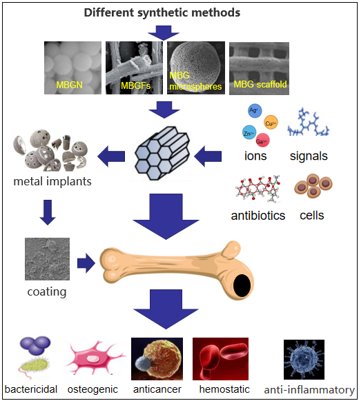
骨填充和骨替代生物材料的研究与开发是骨修复领域重要的研究方向之一。介孔生物活性玻璃(MBG)因其具有良好的生物活性以及可调节的孔径和有序的介孔结构,将在骨修复再生中发挥重要作用。通过不同的制备加工方法能得到纤维、支架、中空结构或纳米颗粒结构的MBG;大量研究表明,在MBG中掺杂少量的治疗性无机离子,能赋予它们更多的生物学特性,包括成骨、抗菌、抗炎、止血或抗癌特性;而且无机离子掺杂的MBG作为支架或纳米颗粒加工后,仍然具有出色的生物活性反应。此外,通过在介孔结构内负载生物活性分子、治疗药物和干细胞可进一步改善MBG的性能。本文介绍了近年来MBG的合成、金属离子掺杂MBG的抗菌性能以及MBG在其他方面的应用进展。

The research and development of bone filling and bone substitute biomaterials is one of the important research directions in the field of bone repair. Mesoporous bioactive glass (MBG) will play an important role in bone repair and regeneration because of its good bioactivity, adjustable pore size and ordered mesoporous structure. MBG with fiber, scaffold, hollow structure or nano-particle structure can be obtained by different preparation and processing methods. Many studies have shown that the incorporation of a small amount of therapeutic inorganic ions into MBG can endow them with more biological properties, including osteogenic, antibacterial, anti-inflammatory, hemostatic or anti-cancer properties. Moreover, MBG doped with inorganic ions still has excellent bioactivity after being processed as scaffolds or nanoparticles. In addition, the performance of MBG can be further improved by loading bioactive molecules, therapeutic drugs and stem cells into the mesoporous structure. In this paper, the synthesis of MBG, the antibacterial properties of metal ion-doped MBG and the application of MBG in other fields are reviewed.

Contents

1 Introduction

2 MBG synthesis

2.1 Preparation of MBG nanoparticles

2.2 Preparation of MBG fiber (MBGF)

2.3 Preparation of MBG microspheres

2.4 Preparation of MBG scaffold

3 MBG used as antimicrobial carrier

3.1 MBG as an antibiotic carrier

3.2 MBG used as antibacterial ion carrier

3.3 MBG doped with other antiseptic

4 MBG in other applications

4.1 MBG used in hemostasis

4.2 MBG used in anti-inflammation

4.3 MBG used in anti-cancer

4.4 MBG as coating material

5 Conclusion and prospect

()
图1 BG/Gel-1(a 和 b)、 BG/Gel-2(c 和 d)和 BG/Gel-3(e 和 f)的 SEM 图和纳米纤维直径分布[56]
Fig. 1 SEM micrographs and nanofiber diameter distributions of BG/Gel-1 (a and b), BG/Gel-2 (c and d) and BG/Gel-3 (e and f) [56]
图2 (a)Ag@pMBG 制备的示意图,即通过邻苯二酚基团的氧化还原活性在 MBG 介孔通道的内表面和外表面原位还原 Ag+ 至 Ag 纳米颗粒。(b)用选择性激光烧结制备多孔复合支架的示意图。典型多孔支架(PLLA-PGA/ Ag@pMBG 和 PLLA-PGA/MBG)的宏观形貌(光学图像)表现出均匀的孔和支架结构[83]
Fig. 2 (a) Schematic of Ag@pMBG preparation, in situ reduction of Ag+ to Ag nanoparticles on the inner and outer surfaces of mesoporous channel of MBG by redox activity of catechol groups. (b) Schematic for preparation of porous composite scaffolds by selective laser sintering. The macroscopic morphology (optical images) of the typical porous scaffolds (PLLA-PGA/Ag@pMBG and PLLA-PGA/MBG) demonstrated a uniform pore and strut structure[83]
图3 合成MBG(a,c)和Zn-MBG(b,d)的SEM图[91]
Fig. 3 Morphology of synthesized MBG (a, c) and Zn-MBG (b, d) particles obtained by SEM [91]
图4 MBG的SEM图和粒度分布[114]
Fig. 4 SEM images and particle size distribution of MBG[114]
图5 Ga-MBG的SEM图[120]:(a)Ga1,(b)Ga3,(c)Ga5
Fig. 5 SEM of Ga-MBG [120]: (a) Ga1, (b) Ga3 and(c)Ga5
表1 离子掺杂MBG抗菌的主要研究汇总
Table 1 Summary of major studies on ion-doped MBG antibacterial agents
Ion(s) doped Composition Synthesis method Biocompatibility Bacterial
species
Antibacterial properties Refs
Ag+/
Ag
PLLA-PGA/xAg@pMBG
(x=0/2.0/4.0/6.0/ 8.0mol%)
Sol-Gel The cells cultured on the scaffold with MBG exhibited a flatter morphology, indicating better cytocompatibility.
the degradation of MBG releases active elements (silicon and calcium) that could induce osteoblast differentiation.
E. coli PLLA-PGA/MBG scaffolds had no antibacterial activity;
The bacteriostasis rates of composite scaffolds loaded with 2AG@PMBG and AG@PMBG were 80.0% and 83.0% ,respectively;
The bacteriostasis rate of composite scaffolds loaded with 6AG@PMBG and 8Ag@pMBG was more than 99.00%;
83
Zn2+ 70SiO2-(30-x)CaO-xZnO
(x=0/2.0/4.0mol%)
Sol-Gel All samples show very high levels of cell mitochondrial activity (> 80.0% of the reference control).
The results indicate that all glasses are not cytotoxic (viability is always above 80.0%, indicates that the material should be considered as not cytotoxic) and favorable for cell proliferation.
E. coli
S.aureus
When the ratio of the extract to the bacterial mixture was 1ml: 0.5 ml, the bacteriostasis rates of 2Zn-MBG to both bacteria were 100.0%, and the bacteriostasis rates of 4Zn-MBG to E. coli and S. aureus were 65.0% and 70.0%, respectively;
When the ratio was 1ml: 0.2 ml, the inhibition rates of 2Zn-MBG to both bacteria were 100%, while those of 4Zn-MBG to E. coli and S. aureus were 80.0% and 85.0%,respectively;
92
Cu2+ 45SiO2-6P2O5-24.5CaO-(24.5-x)Na2O (x=0/0.5/1.5/2.5
mol%)
Sol-Gel Ionic radii variations can influence the dissolution of calcium and phosphate, so apatite growth was gradually increased by addition of copper in BG system.
BG and copper incorporated BG showed almost similar bioactivity as well as exhibiting same apatite growth with the additional benefit of copper release.
P.aeruginosa
E. coli
B.subtilis
S. aureus
E.faecalis
C.albicans
Under the treatment of 1.5 cu-mbg and 2.5 cu-mbg, the viable cells of Pseudomonas aeruginosa and E. coli were completely inhibited, and the inhibitory effects on B. subtilis and S. aureus were obviously superior to those of Gram-negative;
CuBGs (20.0 μg/ml) had a rapid inhibitory effect on Gram-positive bacteria such as Enterobacter faecalis, Candida albicans and Staphylococcus aureus, with inhibition rates of 98.5%, 99.0% and 98.5%, respectively;
97
Ce3+ 88.5SiO2-10.1
CaO-1.4Ce2O3
Sol-Gel some apatite particles existed as hollow hemispheres on day 3 and day 7. as the immersion time increased to 14 days, when most of the apatite particles had grown into full spheres.
All samples induced the formation of apatite particles with Ca/P ratio close to 1.67 upon immersion in simulated body fluid (SBF), confirming their good bioactivity.
E. coli
S. aureus
Ce-MBG has antibacterial activity against Gram-positive bacteria and Gram-negative bacteria by producing reactive oxygen species;
When the concentration was 0.01 mg/ml, it had no effect on E. coli, but the survival rate decreased with the increase of MBG concentration;
When the concentration was 10.0 mg/ml, the growth of Staphylococcus aureus was inhibited completely;
113
Ce3+/
Ga3+
60.0SiO2-(40.0-
x)CaO-xGa2O3
(x=0/1.0/3.0/5.0mol%)
Sol-Gel The Ca/P ratio on the MBGNPs surface was close to 1.64, which is similar to the Ca/P ratio in HA.
All Ga-doped MBGNPs showed the formation of a similar type of HA crystals on the surface. Increasing the amount of gallium doping resulted in significant refinement of precipitated HA crystals.
E. coli
S. aureus
Ga-MBG had a lower survival rate than Gram-positive bacteria and Gram-negative;
The inhibition rate of 5Ga-MBG was the highest at 6h;
Ga1 was the strongest at 6h and 24h after Gram-positive bacteria Gram-negative;
The inhibition rates of Ga1, Ga3 and Ga5 MBG on Gram-negative were significantly different at 24 h incubation.
120
图6 多孔支架在体液中浸泡28? d后的SEM图:(a)未使用和(b)使用58S生物玻璃涂层[154]
Fig. 6 SEM images of porous Vitallium after 28?days soaking in SBF: (a) without and (b) with 58S Bioglass coating[154]
[1]
Wang S S, Zhu T T, Wang D P, Zhang M R, Wang X K, Yu Y, Dong H L, Wu G Z, Zhang M L. Front. Bioeng. Biotechnol., 2023, 11: 1081446.
[2]
Croes M, van der Wal B C H, Vogely H C. J. Orthop. Res., 2019, 37(10): 2067.

doi: 10.1002/jor.v37.10     URL    
[3]
Rioja A Y, Daley E L H, Habif J C, Putnam A J, Stegemann J P. Acta Biomater., 2017, 55: 144.

doi: 10.1016/j.actbio.2017.03.050     URL    
[4]
Keating J F, Simpson A H R W, Robinson C M. J. Bone Jt. Surg. Br. Vol., 2005, 87-B(2): 142.
[5]
Annamalai R T, Hong X W, Schott N G, Tiruchinapally G, Levi B, Stegemann J P. Biomaterials, 2019, 208: 32.

doi: 10.1016/j.biomaterials.2019.04.001     URL    
[6]
Campana V, Milano G, Pagano E, Barba M, Cicione C, Salonna G, Lattanzi W, Logroscino G. J. Mater. Sci. Mater. Med., 2014, 25(10): 2445.

doi: 10.1007/s10856-014-5240-2     URL    
[7]
Zhao T C, Chen L, Lin R F, Zhang P F, Lan K, Zhang W, Li X M, Zhao D Y. Acc. Mater. Res., 2020, 1(1): 100.

doi: 10.1021/accountsmr.0c00028     URL    
[8]
Jones J R. Acta Biomater., 2013, 9(1): 4457.

doi: 10.1016/j.actbio.2012.08.023     URL    
[9]
Li R, Clark A E, Hench L L. J. Appl. Biomater., 1991, 2(4): 231.

doi: 10.1002/jab.v2:4     URL    
[10]
Pereira M M, Clark A E, Hench L L. J. Biomed. Mater. Res., 1994, 28(6): 693.

doi: 10.1002/jbm.v28:6     URL    
[11]
Ladrón de Guevara-Fern S. Biomaterials, 2003, 24(22): 4037.

doi: 10.1016/S0142-9612(03)00279-5     URL    
[12]
Yan X X, Yu C Z, Zhou X F, Tang J W, Zhao D Y. Angew. Chem. Int. Ed., 2004, 43(44): 5980.

doi: 10.1002/anie.v43:44     URL    
[13]
López-Noriega A, Arcos D, Izquierdo-Barba I, Sakamoto Y, Terasaki O, Vallet-Regí M. Chem. Mater., 2006, 18(13): 3137.

doi: 10.1021/cm060488o     URL    
[14]
Yanagisawa T, Shimizu T, Kuroda K, Kato C. Bull. Chem. Soc. Jpn., 1990, 63(4): 988.

doi: 10.1246/bcsj.63.988     URL    
[15]
Horcajada P, Rámila A, Boulahya K, González-Calbet J, Vallet-Regí M. Solid State Sci., 2004, 6(11): 1295.

doi: 10.1016/j.solidstatesciences.2004.07.026     URL    
[16]
Montazerian M, Zanotto E D. J. Mater. Sci., 2017, 52(15): 8695.

doi: 10.1007/s10853-017-0804-4     URL    
[17]
Hsu F Y, Weng R C, Lin H M, Lin Y H, Lu M R, Yu J L, Hsu H W. Microporous Mesoporous Mater., 2015, 212: 56.

doi: 10.1016/j.micromeso.2015.03.027     URL    
[18]
Mohan Raj R, Priya P, Raj V. J. Mech. Behav. Biomed. Mater., 2018, 82: 299.

doi: 10.1016/j.jmbbm.2017.12.033     URL    
[19]
Zhu M, Li K, Zhu Y F, Zhang J H, Ye X J. Acta Biomater., 2015, 16: 145.

doi: 10.1016/j.actbio.2015.01.034     URL    
[20]
Liu F, Bai L B, Zhang H L, Song H Z, Hu L D, Wu Y G, Ba X W. ACS Appl. Mater. Interfaces, 2017, 9(37): 31626.

doi: 10.1021/acsami.7b10867     URL    
[21]
Moghanian A, Firoozi S, Tahriri M, Sedghi A. Mat Sci Eng C-Mater, 2018, 91: 345.
[22]
Ashour A A, Felemban M F, Felemban N H, Enan E T, Basha S, Hassan M M, Gad El-Rab S M F. Antibiotics (Basel), 2022, 11(6): 2079.
[23]
Zheng K, Fan Y Q, Torre E, Balasubramanian P, Taccardi N, Cassinelli C, Morra M, Iviglia G, Boccaccini A R. Part. Part. Syst. Charact., 2020, 37(7): 2000054.
[24]
Wu C T, Chang J. J. Control. Release, 2014, 193: 282.

doi: 10.1016/j.jconrel.2014.04.026     URL    
[25]
Li C, Liu L D, Zhou Z Y, Liu T Y, Zhang S Y, Lu A X. Ceram. Int., 2022, 48(9): 12430.

doi: 10.1016/j.ceramint.2022.01.108     URL    
[26]
Ebrahimi M, Manafi S, Sharifianjazi F. J. Non Cryst. Solids, 2023, 606: 122189.

doi: 10.1016/j.jnoncrysol.2023.122189     URL    
[27]
Shih S J, Lin Y C, Valentino Posma Panjaitan L, Rahayu Meyla Sari D. Materials, 2016, 9(1): 58.

doi: 10.3390/ma9010058     URL    
[28]
Xia W, Chang J. Mater. Lett., 2007, 61(14/15): 3251.

doi: 10.1016/j.matlet.2006.11.048     URL    
[39]
Yan X X, Deng H X, Huang X H, Lu G Q, Qiao S Z, Zhao D Y, Yu C Z. J. Non Cryst. Solids, 2005, 351(40/42): 3209.

doi: 10.1016/j.jnoncrysol.2005.08.024     URL    
[30]
Wu C T, Chang J. Interface Focus., 2012, 2(3): 292.

doi: 10.1098/rsfs.2011.0121     URL    
[31]
Wen C L, Qian J M, Xiao L, Luo L J, Zheng J R, Xie M J, Tao J, Wu X H, Sa B S, Luo K. Ceram. Int., 2022, 48(22): 33781.

doi: 10.1016/j.ceramint.2022.07.324     URL    
[32]
Zhao Z J, Liao Y D, Kong D X, Chen X T, Jiao Y F, Zhang J, Gao P, Zhang X C, Yu W F. Mater. Lett., 2022, 306: 130891.

doi: 10.1016/j.matlet.2021.130891     URL    
[33]
Bokov D, Turki Jalil A, Chupradit S, Suksatan W, Javed Ansari M, Shewael I H, Valiev G H, Kianfar E. Adv. Mater. Sci. Eng., 2021, 2021: 5102014.
[34]
Simila H O, Boccaccini A R. Front. Bioeng. Biotechnol., 2023, 11: 1065597.

doi: 10.3389/fbioe.2023.1065597     URL    
[35]
Zhang W, Zhang X N, Zhou Y, Zhang Y. Ceram. Int., 2023, 49(14): 22924.

doi: 10.1016/j.ceramint.2023.04.117     URL    
[36]
Luo H L, Xiao J, Peng M X, Zhang Q C, Yang Z W, Si H J, Wan Y Z. J. Non Cryst. Solids, 2020, 532: 119856.

doi: 10.1016/j.jnoncrysol.2019.119856     URL    
[37]
Hong Y L, Chen X S, Jing X B, Fan H S, Guo B, Gu Z W, Zhang X D. Adv. Mater., 2010, 22(6): 754.

doi: 10.1002/adma.v22:6     URL    
[38]
Qin X, Cao R, Zheng J J, Shi G J, Ji L J, Zhu A P, Yao H. RSC Adv., 2020, 10(73): 44835.

doi: 10.1039/D0RA08656H     URL    
[39]
Lu H H, Zheng K, Boccaccini A R, Liverani L. Mater. Lett., 2023, 334: 133712.

doi: 10.1016/j.matlet.2022.133712     URL    
[40]
Izquierdo-Barba I, Vallet-Regí M. Biomed. Glasses, 2015, 1(1): 140.
[41]
Wu C T, Zhang Y F, Ke X B, Xie Y X, Zhu H Y, Crawford R, Xiao Y. J. Biomed. Mater. Res. Part A, 2010, 95A(2): 476.

doi: 10.1002/jbm.a.v95a:2     URL    
[42]
Yun H S, Kim S E, Hyun Y T. Mater. Chem. Phys., 2009, 115(2/3): 670.

doi: 10.1016/j.matchemphys.2009.02.001     URL    
[43]
Duan H B, Diao J J, Zhao N R, Ma Y J. Mater. Lett., 2016, 167: 201.

doi: 10.1016/j.matlet.2015.12.162     URL    
[44]
Wu Z Y, Hill R G, Yue S, Nightingale D, Lee P D, Jones J R. Acta Biomater., 2011, 7(4): 1807.

doi: 10.1016/j.actbio.2010.11.041     URL    
[45]
Najafi N, Heuzey M C, Carreau P J, Therriault D, Park C B. Rheol. Acta, 2014, 53(10): 779.

doi: 10.1007/s00397-014-0801-3     URL    
[46]
Zhou Y J, Tian Y R, Peng X W. Polymers, 2023, 15(2): 402.

doi: 10.3390/polym15020402     URL    
[47]
Tainio J, Paakinaho K, Ahola N, Hannula M, Hyttinen J, Kellomäki M, Massera J. Materials, 2017, 10(11): 1274.

doi: 10.3390/ma10111274     URL    
[48]
Song C B, Zhang J P, Li S, Yang S B, Lu E Y, Xi Z H, Cen L, Zhao L, Yuan W K. Chin. J. Chem. Eng., 2021, 29: 426.

doi: 10.1016/j.cjche.2020.07.063     URL    
[49]
Li S, Song C B, Yang S B, Yu W J, Zhang W Q, Zhang G H, Xi Z H, Lu E Y. Acta Biomater., 2019, 94: 253.

doi: 10.1016/j.actbio.2019.05.066     URL    
[50]
Guo R, Hou X H, Zhao D K, Wang H L, Shi C X, Zhou Y. J. Non Cryst. Solids, 2022, 583: 121481.

doi: 10.1016/j.jnoncrysol.2022.121481     URL    
[51]
Kandasamy S, Naveen R. J. Food Process. Eng., 2022, 45(8): e14059.

doi: 10.1111/jfpe.v45.8     URL    
[52]
Santos D, Martins T, Carvalho S, Pereira M, Houmard M, Nunes E. Mater. Lett., 2019, 256: 126647.

doi: 10.1016/j.matlet.2019.126647     URL    
[53]
Atkinson I, Seciu-Grama A M, Petrescu S, Culita D, Mocioiu O C, Voicescu M, Mitran R A, Lincu D, Prelipcean A M, Craciunescu O. Pharmaceutics, 2022, 14(6): 1169.

doi: 10.3390/pharmaceutics14061169     URL    
[54]
Xu J, Zhu X Z, Xu L H, Kan C X, Shi D N. Nanoscale, 2023, 15(4): 1687.

doi: 10.1039/D2NR05667D     URL    
[55]
Wu C T, Xia L G, Han P P, Mao L X, Wang J C, Zhai D, Fang B, Chang J, Xiao Y. ACS Appl. Mater. Interfaces, 2016, 8(18): 11342.

doi: 10.1021/acsami.6b03100     URL    
[56]
Luo H L, Zhang Y, Wang Z R, Yang Z W, Tu J P, Liu Z H, Yao F L, Xiong G Y, Wan Y Z. Chem. Eng. J., 2017, 326: 210.

doi: 10.1016/j.cej.2017.05.115     URL    
[57]
Karakurt I, Lin L W. Curr. Opin. Chem. Eng., 2020, 28: 134.

doi: 10.1016/j.coche.2020.04.001     URL    
[58]
Sun K, Li R X, Jiang W X, Sun Y F, Li H. Biochem. Biophys. Res. Commun., 2016, 477(4): 1085.

doi: 10.1016/j.bbrc.2016.07.050     URL    
[59]
Du X Y, Wei D X, Huang L, Zhu M, Zhang Y P, Zhu Y F. Mater. Sci. Eng. C, 2019, 103: 109731.

doi: 10.1016/j.msec.2019.05.016     URL    
[60]
Chen Y, Chen L, Wang Y T, Lin K L, Liu J Q. Compos. Part B Eng., 2022, 230: 109550.

doi: 10.1016/j.compositesb.2021.109550     URL    
[61]
Liao M H, Zhu S L, Guo A J, Han X Y, Li Q T, Chen Y, Liu Y W, Chen D F, Chen X F, Mo S X, Cao X D. Compos. Part B Eng., 2023, 254: 110582.

doi: 10.1016/j.compositesb.2023.110582     URL    
[62]
Perry K I, Hanssen A D. J. Am. Acad. Orthop. Surg., 2017, 25(1): S4.

doi: 10.5435/JAAOS-D-16-00634     URL    
[63]
Griffith M, Islam M M, Edin J, Papapavlou G, Buznyk O, Patra H K. Front. Bioeng. Biotechnol., 2016, 4: 71.
[64]
Kaya S, Cresswell M, Boccaccini A R. Mater. Sci. Eng. C, 2018, 83: 99.

doi: 10.1016/j.msec.2017.11.003     URL    
[65]
Pajares-Chamorro N, Wagley Y, Hammer N, Hankenson K, Chatzistavrou X. J Am Ceram Soc., 2022, 105(3): 1778.

doi: 10.1111/jace.v105.3     URL    
[66]
Anand A, Das P, Nandi S K, Kundu B. Ceram. Int., 2020, 46(4): 5477.

doi: 10.1016/j.ceramint.2019.10.264     URL    
[67]
Zhang W, Zhu K R, Ren W X, He H L, Liang H C, Zhai Y P, Li W. Adv. Mater. Interfaces, 2022, 9(3): 2101528.
[68]
Zhu Y F, Kaskel S. Microporous Mesoporous Mater., 2009, 118(1/3): 176.

doi: 10.1016/j.micromeso.2008.08.046     URL    
[69]
Liang M H, Liu Q. J. Healthc. Eng., 2022, 2022: 2114661.
[70]
Xue J M, Shi M. J. Control. Release, 2004, 98(2): 209.

doi: 10.1016/j.jconrel.2004.04.023     URL    
[71]
Xia W, Chang J. J. Control. Release, 2006, 110(3): 522.

doi: 10.1016/j.jconrel.2005.11.002     URL    
[72]
Sánchez-Salcedo S, Heras C, Lozano D, Vallet-Regí M, Salinas A J. Acta Biomater., 2023, 166: 655.

doi: 10.1016/j.actbio.2023.04.046     URL    
[73]
Khanmohammadi S, Aghajani H, Farrokhi-Rad M. Ceram. Int., 2022, 48(14): 20176.

doi: 10.1016/j.ceramint.2022.03.296     URL    
[74]
Patil A, Banerji R, Kanojiya P, Koratkar S, Saroj S. Expert Rev. Anti Infect. Ther., 2021, 19(7): 845.

doi: 10.1080/14787210.2021.1858800     URL    
[75]
Slavin Y N, Asnis J, Häfeli U O, Bach H. J. Nanobiotechnol., 2017, 15(1): 65.

doi: 10.1186/s12951-017-0308-z     URL    
[76]
Wang X J, Li W. Nanotechnology, 2016, 27(22): 225102.

doi: 10.1088/0957-4484/27/22/225102     URL    
[77]
Barrioni B R, Oliveira A C, de Fátima Leite M, de Magalhães Pereira M. J. Mater. Sci., 2017, 52(15): 8904.

doi: 10.1007/s10853-017-0944-6     URL    
[78]
Özarslan A C, Yücel S. Ceram Int, 2023, 49(9): 13940.

doi: 10.1016/j.ceramint.2022.12.276     URL    
[79]
Bouhazma S, Chajri S, Khaldi M, Sadiki M, Barkai H, Elabed S, Ibnsouda Koraichi S, El Bali B, Lachkar M. IOP Conf. Ser.: Mater. Sci. Eng., 2017, 186: 012022.

doi: 10.1088/1757-899X/186/1/012022     URL    
[80]
Raimondi S, Zambon A, Ranieri R, Fraulini F, Amaretti A, Rossi M, Lusvardi G. J. Biomed. Mater. Res. Part A, 2022, 110(2): 504.

doi: 10.1002/jbm.a.v110.2     URL    
[81]
Carta D, Jones J R, Lin S, Poologasundarampillai G, Newport R J, Pickup D M. Int. J. Appl. Glass Sci., 2017, 8(4): 364.

doi: 10.1111/ijag.2017.8.issue-4     URL    
[82]
Zheng K, Balasubramanian P, Paterson T E, Stein R, MacNeil S, Fiorilli S, Vitale-Brovarone C, Shepherd J, Boccaccini A R. Mater. Sci. Eng. C, 2019, 103: 109764.

doi: 10.1016/j.msec.2019.109764     URL    
[83]
Shuai C J, Xu Y, Feng P, Wang G Y, Xiong S X, Peng S P. Chem. Eng. J., 2019, 374: 304.

doi: 10.1016/j.cej.2019.03.273     URL    
[84]
Huang Y C, Yang T Y, Chen B X, Kung J C, Shih C J. Pharmaceuticals, 2021, 14(11): 1094.

doi: 10.3390/ph14111094     URL    
[85]
Balasubramanian P, Strobel L A, Kneser U, Boccaccini A R. Biomed. Glasses, 2015, 1(1): 51.
[86]
Pasquet J, Chevalier Y, Couval E, Bouvier D, Noizet G, Morlière C, Bolzinger M A. Int. J. Pharm., 2014, 460(1/2): 92.

doi: 10.1016/j.ijpharm.2013.10.031     URL    
[87]
Webster T J, Seil I. Int. J. Nanomed., 2012: 2767.
[88]
Zhang L L, Jiang Y H, Ding Y L, Povey M, York D. J. Nanopart. Res., 2007, 9(3): 479.

doi: 10.1007/s11051-006-9150-1     URL    
[89]
Stankovic A, Dimitrijevic S, Uskokovic D. Colloids Surf B Biointerfaces, 2013, 102: 21.

doi: 10.1016/j.colsurfb.2012.07.033     URL    
[90]
Leung Y H, Chan C N, Ng A M C, Chan H T, Chiang M L, Djurišić A B, Ng Y H, Jim W Y, Guo M Y, Leung F C, Chan W K, Au D T W. Nanotechnology, 2012, 23(47): 475703.

doi: 10.1088/0957-4484/23/47/475703     URL    
[91]
Nescakova Z, Zheng K, Liverani L, Nawaz Q, Galuskova D, Kankova H, Michalek M, Galusek D, Boccaccini A R. Bioact Mater, 2019, 4: 86.
[92]
Wajda A, Goldmann W H, Detsch R, Boccaccini A R, Sitarz M. J. Non Cryst. Solids, 2019, 511: 86.

doi: 10.1016/j.jnoncrysol.2018.12.040     URL    
[93]
Grass G, Rensing C, Solioz M. Appl. Environ. Microbiol., 2011, 77(5): 1541.

doi: 10.1128/AEM.02766-10     URL    
[94]
Markovic D, Deeks C, Nunney T, Radovanovic Z, Radoicic M, Saponjic Z, Radetic M. Carbohydr Polym, 2018, 200: 173.

doi: 10.1016/j.carbpol.2018.08.001     URL    
[95]
Ren G G, Hu D W, Cheng E W C, Vargas-Reus M A, Reip P, Allaker R P. Int. J. Antimicrob. Agents, 2009, 33(6): 587.

doi: 10.1016/j.ijantimicag.2008.12.004     URL    
[96]
Wu C T, Zhou Y H, Xu M C, Han P P, Chen L, Chang J, Xiao Y. Biomaterials, 2013, 34(2): 422.

doi: 10.1016/j.biomaterials.2012.09.066     URL    
[97]
Chitra S, Bargavi P, Balasubramaniam M, Chandran R R, Balakumar S. Mater. Sci. Eng. C, 2020, 109: 110598.

doi: 10.1016/j.msec.2019.110598     URL    
[98]
Yang Y W, Lu C F, Yang M L, Wang D S, Peng S P, Tian Z J, Shuai C J. Mater. Chem. Front., 2021, 5(19): 7228.

doi: 10.1039/D1QM01028J     URL    
[99]
Hu S, Chang J, Liu M Q, Ning C Q. J. Mater. Sci. Mater. Med., 2009, 20(1): 281.

doi: 10.1007/s10856-008-3564-5     URL    
[100]
Mortazavi V, Nahrkhalaji M M, Fathi M H, Mousavi S B, Esfahani B N. J. Biomed. Mater. Res. Part A, 2010, 94A(1): 160.

doi: 10.1002/jbm.a.v94a:1     URL    
[101]
Cha S H, Hong J, McGuffie M, Yeom B, VanEpps J S, Kotov N A. ACS Nano, 2015, 9(9): 9097.

doi: 10.1021/acsnano.5b03247     URL    
[102]
Sharan R, Chhibber S, Attri S, Reed R H. Antonie Van Leeuwenhoek, 2010, 97(1): 91.

doi: 10.1007/s10482-009-9395-7     URL    
[103]
Tian W X, Yu S, Ibrahim M, Almonaofy A W, He L, Hui Q, Bo Z, Li B, Xie G L. J. Microbiol., 2012, 50(4): 586.

doi: 10.1007/s12275-012-2067-8     URL    
[104]
Studer A M, Limbach L K, Van Duc L, Krumeich F, Athanassiou E K, Gerber L C, Moch H, Stark W J. Toxicol. Lett., 2010, 197(3): 169.

doi: 10.1016/j.toxlet.2010.05.012     URL    
[105]
Lv Y R, Li L, Yin P, Lei T. Dalton Trans., 2020, 49(15): 4699.

doi: 10.1039/D0DT00201A     URL    
[106]
Fan X Z, Yahia L, Sacher E. Biology, 2021, 10(2): 137.

doi: 10.3390/biology10020137     URL    
[107]
Raffi M, Mehrwan S, Bhatti T M, Akhter J I, Hameed A, Yawar W, ul Hasan M M. Ann. Microbiol., 2010, 60(1): 75.

doi: 10.1007/s13213-010-0015-6     URL    
[108]
Scheidegger D, Sparkes B G, Lüscher N, Schoenenberger G A, Allgöwer M. Burns, 1992, 18(4): 296.

doi: 10.1016/0305-4179(92)90150-S     URL    
[109]
Kistler D, Hafemann B, Schoenenberger G A, Hettich R. Eur. Surg. Res., 1990, 22(5): 283.

doi: 10.1159/000129114     URL    
[110]
Chen B H, Suresh Babu K, Anandkumar M, Tsai T Y, Kao T H, Stephen Inbaraj B. Int. J. Nanomed., 2014: 5515.
[111]
AshaRani P V, Low Kah Mun G, Hande M P, Valiyaveettil S. ACS Nano, 2009, 3(2): 279.

doi: 10.1021/nn800596w     URL    
[112]
Ingle A P, Duran N, Rai M. Appl. Microbiol. Biotechnol., 2014, 98(3): 1001.

doi: 10.1007/s00253-013-5422-8     URL    
[113]
Goh Y F, Alshemary A Z, Akram M, Abdul Kadir M R, Hussain R. Ceram. Int., 2014, 40(1): 729.

doi: 10.1016/j.ceramint.2013.06.062     URL    
[114]
Kurtuldu F, Kaňková H, Beltrán A M, Liverani L, Galusek D, Boccaccini A R. Mater. Today Bio, 2021, 12: 100150.
[115]
Zhang M Z, Zhang C, Zhai X Y, Luo F, Du Y P, Yan C H. Sci. China Mater., 2019, 62(11): 1727.

doi: 10.1007/s40843-019-9471-7    
[116]
Westhauser F, Rehder F, Decker S, Kunisch E, Moghaddam A, Zheng K, Boccaccini A R. Biomed. Mater., 2021, 16(3): 035028.

doi: 10.1088/1748-605X/abcf5f    
[117]
Bullen J J, Rogers H J, Spalding P B, Ward C G. FEMS Immunol. Med. Microbiol., 2005, 43(3): 325.

doi: 10.1016/j.femsim.2004.11.010     URL    
[118]
Chitambar C R. Biochim. Biophys. Acta BBA Mol. Cell Res., 2016, 1863(8): 2044.
[119]
Pourshahrestani S, Zeimaran E, Adib Kadri N, Gargiulo N, Samuel S, Naveen S V, Kamarul T, Towler M R. J. Mater. Chem. B, 2016, 4(1): 71.

doi: 10.1039/C5TB02062J     URL    
[120]
Kurtuldu F, Mutlu N, Michálek M, Zheng K, Masar M, Liverani L, Chen S, Galusek D, Boccaccini A R. Mater. Sci. Eng. C, 2021, 124: 112050.

doi: 10.1016/j.msec.2021.112050     URL    
[121]
Łapa A, Cresswell M, Campbell I, Jackson P, Goldmann W H, Detsch R, Boccaccini A R. Adv. Eng. Mater., 2020, 22(9): 1901577.

doi: 10.1002/adem.v22.9     URL    
[122]
Westhauser F, Wilkesmann S, Nawaz Q, Schmitz S I, Moghaddam A, Boccaccini A R. J. Biomed. Mater. Res. Part A, 2020, 108(9): 1806.

doi: 10.1002/jbm.a.v108.9     URL    
[123]
Ye Q, Chen W, Huang H, Tang Y Q, Wang W X, Meng F R, Wang H L, Zheng Y S. Appl. Microbiol. Biotechnol., 2020, 104(12): 5213.

doi: 10.1007/s00253-020-10600-4    
[124]
Baheiraei N, Eyni H, Bakhshi B, Najafloo R, Rabiee N. Sci. Rep., 2021, 11: 8745.

doi: 10.1038/s41598-021-88058-1    
[125]
Shih S J, Chen C Y, Lin Y C, Lee J C, Chung R J. Adv. Powder Technol., 2016, 27(3): 1013.

doi: 10.1016/j.apt.2016.04.016     URL    
[126]
Tseng C F, Fei Y C, Chou Y J. J. Non Cryst. Solids, 2020, 549: 120336.

doi: 10.1016/j.jnoncrysol.2020.120336     URL    
[127]
Liu J, Rawlinson S C F, Hill R G, Fortune F. Dent. Mater., 2016, 32(3): 412.

doi: 10.1016/j.dental.2015.12.013     URL    
[128]
Akhavan O, Ghaderi E. ACS Nano, 2010, 4(10): 5731.

doi: 10.1021/nn101390x     URL    
[129]
Krishnamoorthy K, Umasuthan N, Mohan R, Lee J, Kim S J. Sci. Adv. Mater., 2012, 4(11): 1111.

doi: 10.1166/sam.2012.1402     URL    
[130]
Bannow B S, Konkle B A. Blood, 2023, 142(9): 761.
[131]
Pourshahrestani S, Zeimaran E, Kadri N A, Gargiulo N, Jindal H M, Naveen S V, Sekaran S D, Kamarul T, Towler M R. ACS Appl. Mater. Interfaces, 2017, 9(37): 31381.

doi: 10.1021/acsami.7b07769     URL    
[132]
Salinas A J, Shruti S, Malavasi G, Menabue L, Vallet-Regí M. Acta Biomater., 2011, 7(9): 3452.

doi: 10.1016/j.actbio.2011.05.033     URL    
[133]
Roy P, Saha R, Chakraborty J. Ceram. Int., 2023, 49(4): 6389.

doi: 10.1016/j.ceramint.2022.10.114     URL    
[134]
Liu J X, Zhou X, Zhang Y, Zhu W, Wang A P, Xu M J, Zhuang S X. Mater. Today Chem., 2022, 23: 100735.
[135]
Loi F, Córdova L A, Pajarinen J, Lin T H, Yao Z Y, Goodman S B. Bone, 2016, 86: 119.

doi: 10.1016/j.bone.2016.02.020     URL    
[136]
Goodman S B, Maruyama M. J. Inflamm. Res., 2020, 13: 913.

doi: 10.2147/JIR.S281941     URL    
[137]
Newman H, Shih Y V, Varghese S. Biomaterials, 2021, 277: 121114.

doi: 10.1016/j.biomaterials.2021.121114     URL    
[138]
Mazzaferro S, De Martini N, Rotondi S, Tartaglione L, Ureña-Torres P, Bover J, Pasquali M. Clin. Chim. Acta, 2020, 506: 236.

doi: 10.1016/j.cca.2020.03.040     URL    
[139]
Mazzaferro S, Bagordo D, De Martini N, Pasquali M, Rotondi S, Tartaglione L, Stenvinkel P, Group T E E C M W. Calcif. Tissue Int., 2021, 108(4): 452.

doi: 10.1007/s00223-020-00794-0    
[140]
Maruyama M, Rhee C, Utsunomiya T, Zhang N, Ueno M, Yao Z Y, Goodman S B. Front. Endocrinol., 2020, 11: 386.

doi: 10.3389/fendo.2020.00386     URL    
[141]
Mo Y F, Zhao F J, Lin Z F, Cao X D, Chen D F, Chen X F. Biomater. Sci., 2022, 10(7): 1697.

doi: 10.1039/D1BM01842F     URL    
[142]
Sun H S, Zheng K, Zhou T, Boccaccini A R. Pharmaceutics, 2021, 13(12): 2124.

doi: 10.3390/pharmaceutics13122124     URL    
[143]
Zhang Y, Wu Y J, Qiao X Y, Lin T, Wang Y C, Wang M. Front. Mater., 2022, 9: 990931.

doi: 10.3389/fmats.2022.990931     URL    
[144]
Yu H H, Liu H F, Shen Y, Ao Q. Front. Bioeng. Biotechnol., 2023, 11: 1096525.

doi: 10.3389/fbioe.2023.1096525     URL    
[145]
Wang H. Acc. Mater. Res., 2020, 1(3): 172.

doi: 10.1021/accountsmr.0c00079     URL    
[146]
Taşar C, Ercan B. Ceram. Int., 2023, 49(8): 12925.

doi: 10.1016/j.ceramint.2022.12.164     URL    
[147]
Kermani F, Vojdani-Saghir A, Mollazadeh Beidokhti S, Nazarnezhad S, Mollaei Z, Hamzehlou S, El-Fiqi A, Baino F, Kargozar S. Transl. Oncol., 2022, 20: 101397.

doi: 10.1016/j.tranon.2022.101397     URL    
[148]
Iaquinta M R, Mazzoni E, Bononi I, Rotondo J C, Mazziotta C, Montesi M, Sprio S, Tampieri A, Tognon M, Martini F. Front. Cell Dev. Biol., 2019, 7: 268.

doi: 10.3389/fcell.2019.00268     URL    
[149]
Bekmurzayeva A, Duncanson W J, Azevedo H S, Kanayeva D. Mater. Sci. Eng. C, 2018, 93: 1073.

doi: 10.1016/j.msec.2018.08.049     URL    
[150]
Su Y C, Luo C, Zhang Z H, Hermawan H, Zhu D H, Huang J B, Liang Y H, Li G Y, Ren L Q. J. Mech. Behav. Biomed. Mater., 2018, 77: 90.

doi: 10.1016/j.jmbbm.2017.08.035     URL    
[151]
Asri R I M, Harun W S W, Samykano M, Lah N A C, Ghani S A C, Tarlochan F, Raza M R. Mater. Sci. Eng. C, 2017, 77: 1261.

doi: 10.1016/j.msec.2017.04.102     URL    
[152]
Su Y C, Cockerill I, Zheng Y F, Tang L P, Qin Y X, Zhu D H. Bioact. Mater., 2019, 4: 196.
[153]
Sola A, Bellucci D, Cannillo V, Cattini A. Surf. Eng., 2011, 27(8): 560.

doi: 10.1179/1743294410Y.0000000008     URL    
[154]
Haftbaradaran-Esfahani M, Ahmadian M, Nassajpour-Esfahani A H. Appl. Surf. Sci., 2020, 506: 144959.

doi: 10.1016/j.apsusc.2019.144959     URL    
[155]
Huo S C, Lyu Z C, Su X J, Wang F, Liu J, Liu S, Liu X S, Bao X G, Zhang J, Zheng K, Xu G H. Compos. Part B Eng., 2023, 253: 110521.

doi: 10.1016/j.compositesb.2023.110521     URL    
[156]
Heise S, Höhlinger M, Hernández Y T, Palacio J J P, Rodriquez Ortiz J A, Wagener V, Virtanen S, Boccaccini A R. Electrochim. Acta, 2017, 232: 456.

doi: 10.1016/j.electacta.2017.02.081     URL    
[1] 赵睿, 杨晓, 朱向东, 张兴栋. 微量元素锶掺杂生物材料在骨修复领域的应用[J]. 化学进展, 2021, 33(4): 533-542.
[2] 胡小红 朱旸 高长有. 用于软骨修复的水凝胶*[J]. 化学进展, 2009, 21(10): 2164-2175.