English
新闻公告
More
化学进展 2023, Vol. 35 Issue (12): 1793-1806 DOI: 10.7536/PC230410 前一篇   后一篇

• 综述 •

RIfS干涉基底的制备、应用及展望

苏倩倩1,*(), 孙宇1, 张文文1, 彭正得1, 钱卫平2   

  1. 1 江苏海洋大学药学院 连云港 222005
    2 东南大学生物电子学国家重点实验室 南京 210096
  • 收稿日期:2023-04-10 修回日期:2023-09-10 出版日期:2023-12-24 发布日期:2023-11-30
  • 作者简介:

    苏倩倩 博士,硕士生导师。主要研究方向为生物传感和生化分析。主要研究工作包括:(1)有序多孔纳米材料的制备及性能研究;(2)基于有序多孔纳米材料的生物传感器的研制;(3)生物传感器在药物分析及新药筛选中的应用。作为第一作者在Anal. Chem.ACS Appl. Mater. Interfaces等期刊上发表多篇论文,获PCT国际发明专利授权1项、中国发明专利授权1项、实用新型专利授权1项。2021年获得江苏省“双创博士”人才基金资助。

  • 基金资助:
    数字医学工程全国重点实验室开放研究基金资助课题(2023-K13); 江苏省“双创博士”人才基金项目(JSSCBS20211301); 江苏海洋大学博士科研启动项目(KQ21002)

Preparation, Application and Prospect of RIfS Interference Substrates

Qianqian Su1,*(), Yu Sun1, Wenwen Zhang1, Zhengde Peng1, Weiping Qian2   

  1. 1 Pharmacy School, Jiangsu Ocean University,Lianyungang 222005, China
    2 State Key Laboratory of Bioelectronics, Southeast University, Nanjing 210096, China
  • Received:2023-04-10 Revised:2023-09-10 Online:2023-12-24 Published:2023-11-30
  • Contact: *e-mail: suqianqian@jou.edu.cn
  • Supported by:
    Open Research Fund of State Key Laboratory ofDigital Medical Engineering(2023-K13); Shuangchuang Ph.D award of Jiangsu province(JSSCBS20211301); Doctoral research project of Jiangsu Ocean University(KQ21002)

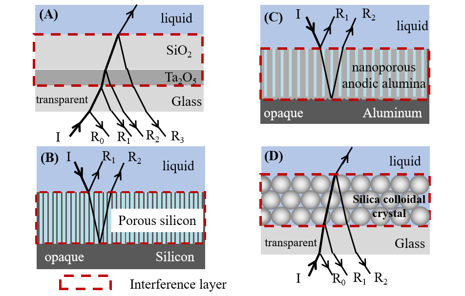
反射干涉光谱(Reflectometric interference spectroscopy,RIfS)是一种利用白光干涉原理来对薄膜光学厚度进行测量的非标记检测技术。干涉基底作为RIfS系统的传感单元,是RIfS技术的核心部分,也是决定RIfS系统性能的关键。目前使用的干涉基底通常被分为两大类:一类是以无机氧化物或聚合物薄膜为代表的平面固体基底,另一类是以多孔硅(pSi)、纳米多孔阳极氧化铝(NAA)以及二氧化硅胶体晶体(SCC)为代表的多孔基底。平面固体基底制备简单且信号稳定,但是检测灵敏度通常比较低;多孔基底具有较大的比表面积,可以捕获更多待测分子,因此与平面固体基底相比检测灵敏度有所提高,并且具有更多的调节空间,非常适合生化传感平台的开发。从pSi到NAA再到SCC,多孔基底制备的可控性及传感性能不断提高,成为RIfS干涉基底的发展方向。本文对RIfS干涉基底发展现状进行了总结和讨论,阐述了基底的常用制备方法,总结了其在生物传感领域代表性的应用,重点讨论了不同基底的优缺点,并对干涉基底未来的发展进行了展望。

Reflectometric interference spectroscopy (RIfS) is a label-free detection technique by measuring the optical thickness of thin films which is based on white light interference. Interference effective substrates, as the sensor unit of the RIfS system, the construction of which is the core part of RIfS technology and the key to determining the performance of the RIfS system. Currently used interference substrates are generally divided into two categories: one is the planar solid substrates represented by inorganic oxides or polymer films, and the other is the porous substrates represented by porous silicon (pSi), nanoporous anodic alumina (NAA) and silica colloidal crystals (SCC). The preparation of planar solid substrate is simple and the signal is stable, but the detection sensitivity is usually low. In comparison with planar solid substrates, a porous substrate can provide a three-dimensional structure with a large specific surface area which will result in increased ligand immobilization density and capture of analyte. Therefore, the detection sensitivity is improved and there is more room for adjustment, which is very suitable for the development of a biochemical sensing platform. From pSi to NAA to SCC, the preparation controllability and sensing performance of porous substrates are continuously improved, which is considered a promising development direction of RIfS interference substrate. Here, the research status of RIfS interference substrates has been summarized and discussed, the common preparation methods of substrates are described, their representative applications in biosensing are summarized, the advantages and disadvantages of different substrates are discussed, and the future development directions of RIfS interference substrates has also been outlined.

Contents

1 Introduction

2 Measurement principles of reflectometric interference spectroscopy

3 Interference substrate

3.1 Planar solid substrate

3.2 Porous silicon substrate

3.3 Nanoporous anodic alumina substrate

3.4 Silica colloidal crystals substrate

4 Conclusion and outlook

()
图1 反射干涉光谱测量原理图
Fig. 1 Schematic diagram of reflectometric interference spectroscopy
图2 多孔硅基底的侧面SEM图[36]
Fig. 2 The SEM image of the side view of porous silicon substrate[36]
表1 基于多孔硅基底的生物传感应用举例[59]
Table 1 Examples of biosensors based on porous silicon substrates[59]
图3 (a) 纳米孔阳极氧化铝基底的结构示意图;(b) 纳米孔阳极氧化铝基底的正面及侧面SEM图(图中标尺分别为400和250 nm);(c) 纳米孔阳极氧化铝基底的制备装置示意图[65]
Fig. 3 (a) Illustrative scheme describing the most representative geometric features of nanoporous anodic alumina (NAA); (b) top and cross-section scanning electron microscopy (SEM) images of NAA (scale bars = 400 and 250 nm, respectively); (c) illustration describing a basic electrochemical anodisation cell used to produce NAA[65]
图4 基于NAA基底的RIfS传感系统的构造示意图[77]
Fig. 4 Schematic diagram of RIfS sensing system based on NAA substrate[77]
表2 胶体晶体的常用制备方法[101]
Table 2 Common preparation methods of colloidal crystals[101]
Method Remarks
Drop casting
(Sedimentation)
Simple but slow process.
Patches of colloidal crystals formed.
Difficult to control exact conditions
Centrifugation Simple and fast process.
Generally big bulky colloidal crystals formed.
Spin-coating Simple and fast process.
Monolayer formation possible. Patches of small coating area of monolayers.
Dip-coating Can control thickness of layers by the speed of withdrawal.
Gradient in layer thickness.
Shear ordering Requires very good control of process parameters.
Slow process. Makes thin films.
Langmuir-Blodgett Monolayer compressed on water surface by mobile arms.
Short range order of closed packed regions within the monolayer.
Monolayer transfer onto substrate can be repeated to deposit multilayers exactly as desired.
Takes time for preparation of equipment and spreading of particles.
Direct assembly on water surface Simple and fast process.
Good two-dimensional closed pack array on water surface.
One monolayer at a time can be transferred.
Can be repeated to deposit multilayers exactly as desired.
Magnetic self-assembly Requires highly-charged monodisperse magnetic colloidal particles.
Self-assembly occurs inside liquid medium.
Can be controlled by external magnetic field.
Vertical deposition Requires very good control of evaporation conditions (i.e., temperature and humidity) for a good deposition.
Slow process (days).
Very good quality of colloidal crystals formed under the proper conditions.
Gradient in the thickness of colloidal crystal formed.
图5 基于硅胶晶体薄膜的反射干涉传感器原理图[100]
Fig. 5 Schematic diagram of the reflectometric interference sensor based on silica colloidal crystal films[100]
[1]
Gauglitz G. Anal. Bioanal. Chem., 2005, 381(1): 141.

pmid: 15700161
[2]
Damborský P, Švitel J, Katrlík J. Essays Biochem., 2016, 60(1): 91.

doi: 10.1042/EBC20150010     pmid: 27365039
[3]
Rau S, Gauglitz G. Anal. Bioanal. Chem., 2012, 402(1): 529.

doi: 10.1007/s00216-011-5470-9     URL    
[4]
Hänel C, Gauglitz G. Anal. Bioanal. Chem., 2002, 372(1): 91.

pmid: 11939217
[5]
Rasooly A, Herold KE. Methods Mol. Biol., 2009, 503: v-ix.
[6]
Langmuir I, Schaefer V J. J. Am. Chem. Soc., 1937, 59(7): 1406.
[7]
Brecht A, Gauglitz G, Polster J. Biosens. Bioelectron., 1993, 8(7/8): 387.

doi: 10.1016/0956-5663(93)80078-4     URL    
[8]
Kraus G, Gauglitz G. FreseniusJ. Anal. Chem., 1992, 344(4/5): 153.
[9]
Weizmann Y, Patolsky F, Willner I. Anal., 2001, 126(9): 1502.

doi: 10.1039/b106613g     URL    
[10]
Leopold N, Busche S, Gauglitz G, Lendl B. Spectrochim. Acta A, 2009, 72(5): 994.

doi: 10.1016/j.saa.2008.12.032     URL    
[11]
Gauglitz G, Brecht A, Kraus G, Mahm W. Sens. Actuat. B, 1993, 11(1/3): 21.

doi: 10.1016/0925-4005(93)85234-2     URL    
[12]
Gauglitz G. Anal. Bioanal. Chem., 2010, 398(6): 2363.

doi: 10.1007/s00216-010-3904-4     pmid: 20623274
[13]
Pacholski C, Sartor M, Sailor M J, Cunin F, Miskelly G M. J. Am. Chem. Soc., 2005, 127(33): 11636.

doi: 10.1021/ja0511671     pmid: 16104739
[14]
Schwartz M P, Alvarez SD, Sailor M J. Anal. Chem., 2007, 79(1): 327.

doi: 10.1021/ac061476p     pmid: 17194157
[15]
Proll G, Steinle L, Pröll F, Kumpf M, Moehrle B, Mehlmann M, Gauglitz G. J. Chromatogr. A, 2007, 1161(1/2): 2.

doi: 10.1016/j.chroma.2007.06.022     URL    
[16]
Busche S, Kasper M, Mutschler T, Leopold N, Gauglitz G. Interaction Behaviour of the Ultramicroporous Polymer Makrolon by Optical Spectroscopic Methods. Eds.: Grundke K, Stamm M, Adler H J. Series: Progress in Colloid and Polymer Science, 2006. 16-22.
[17]
Kasper M, Busche S, Dieterle F, Belge G, Gauglitz G. Meas. Sci. Technol., 2004, 15(3): 540.

doi: 10.1088/0957-0233/15/3/006     URL    
[18]
Pröll F, Möhrle B, Kumpf M, Gauglitz G. Anal. Bioanal. Chem., 2005, 382(8): 1889.

doi: 10.1007/s00216-005-3301-6     URL    
[19]
Brecht A, Ingenhoff J, Gauglitz G. Sens. Actuat. B, 1992, 61-3: 96.
[20]
Zimmermanna R, Osaki T, Gauglitz G, Werner C. Biointerphases, 2007, 2(4): 159.

doi: 10.1116/1.2814066     pmid: 20408653
[21]
Piehler J, Brecht A, Gauglitz G, Zerlin M, Maul C, Thiericke R, Grabley S. Anal. Biochem., 1997, 249(1): 94.

pmid: 9193714
[22]
Yu F, YaoD F, Qian W P. Clin Chem, 2000, 46(9): 1489.

pmid: 10973895
[23]
Lehmann V, Gösele U. Appl. Phys. Lett., 1991, 58(8): 856.

doi: 10.1063/1.104512     URL    
[24]
Herino R, Bomchil G, Barla K, Bertrand C, Ginoux J L. J. Electrochem. Soc., 1987, 134(8): 1994.

doi: 10.1149/1.2100805    
[25]
Li W, Liu Z H, Fontana F, Ding Y P, LiuD F, Hirvonen J T, Santos H A. Adv. Mater., 2018, 30(24): 1703740.

doi: 10.1002/adma.v30.24     URL    
[26]
Lee S, Kang J, KimD. Materials, 2018, 11(12): 2557.

doi: 10.3390/ma11122557     URL    
[27]
Santos H A, Mäkilä E, Airaksinen A, Bimbo L, Hirvonen J. Nanomedicine, 2014, 9(4): 535.

doi: 10.2217/nnm.13.223     URL    
[28]
Low S P, Voelcker N H. Biocompatibility of Porous Silicon. Handbook of Porous Silicon. Cham: Springer, 2014. 1-13.
[29]
Terracciano M, Rea I, Borbone N, Moretta R, Oliviero G, Piccialli G, De Stefano L. Molecules, 2019, 24(12): 2216.

doi: 10.3390/molecules24122216     URL    
[30]
Torres-Costa V, AgullÓ-Rueda F, Martín-Palma R J, Martínez-Duart J M. Opt. Mater., 2005, 27(5): 1084.

doi: 10.1016/j.optmat.2004.08.068     URL    
[31]
Singh S, Sharma S N, Govind, Shivaprasad S M, Lal M, Khan M A. J. Mater. Sci. Mater. Med., 2009, 1:S181.
[32]
Tembe S, Kubal B S, Karve M, D’Souza S F. Anal. Chim. Acta, 2008, 612(2): 212.

doi: 10.1016/j.aca.2008.02.031     URL    
[33]
De Stefano L, Arcari P, Lamberti A, Sanges C, Rotiroti L, Rea I, Rendina I. Sensors, 2007, 7(2): 214.

doi: 10.3390/s7020214     URL    
[34]
Uhlir A Jr. Bell Syst. Tech. J., 1956, 35(2): 333.

doi: 10.1002/bltj.1956.35.issue-2     URL    
[35]
Canham L T. Appl. Phys. Lett., 1990, 57(10): 1046.

doi: 10.1063/1.103561     URL    
[36]
Li Y J, Toan N, Wang Z Q, Bin Samat K F, Ono T. Nanoscale Res. Lett., 2021, 16(1): 1.

doi: 10.1186/s11671-020-03464-0    
[37]
Sailor M J. Porous Silicon in Practice: Preparation, Characterization and Applications, Weinheim:Wiley-VCH, 2012.
[38]
Henstock J R, Ruktanonchai U R, Canham L T, Anderson S I. J. Mater. Sci., 2014, 25(4): 1087.
[39]
Rendina I, Rea I, Rotiroti L, De Stefano L. Phys. E, 2007, 381-2: 188.
[40]
Harraz F A. Actuators B Chem., 2014, 202: 897.

doi: 10.1016/j.snb.2014.06.048     URL    
[41]
Moretta R, Terracciano M, Borbone N, Oliviero G, Schiattarella C, Piccialli G, Falanga A P, Marzano M, Dardano P, De Stefano L, Rea I. Nanomaterials, 2020, 10(11): 2233.

doi: 10.3390/nano10112233     URL    
[42]
Terracciano M, Rea I, De Stefano L, Rendina I, Oliviero G, Nici F, D’Errico S, Piccialli G, Borbone N. Nanoscale Res. Lett., 2014, 9(1): 1.

doi: 10.1186/1556-276X-9-1    
[43]
Krismastuti F S H, Cavallaro A, Prieto-Simon B, Voelcker N H. Adv. Sci., 2016, 3(6): 1500383.

doi: 10.1002/advs.v3.6     URL    
[44]
Ghosh R, Das R, Giri P K. Sens. Actuat. B, 2018, 260: 693.

doi: 10.1016/j.snb.2018.01.099     URL    
[45]
Tieu T, Alba M, Elnathan R, Cifuentes-Rius A, Voelcker N H. Adv. Ther., 2019, 2(1): 1800095.
[46]
Buriak J M, Allen M J. J. Am. Chem. Soc., 1998, 120(6): 1339.

doi: 10.1021/ja9740125     URL    
[47]
Arshavsky-Graham S, Massad-Ivanir N, Segal E, Weiss S. Anal. Chem., 2019, 91(1): 441.

doi: 10.1021/acs.analchem.8b05028     pmid: 30408950
[48]
Dhanekar S, Jain S. Biosens. Bioelectron., 2013, 41: 54.

doi: 10.1016/j.bios.2012.09.045     pmid: 23122704
[49]
Dancil K P S, GreinerD P, Sailor M J. J. Am. Chem. Soc., 1999, 121(34): 7925.

doi: 10.1021/ja991421n     URL    
[50]
Tang Y Y, Li Z, Luo Q H, Liu J Q, Wu J M. Biosens. Bioelectron., 2016, 79: 715.

doi: 10.1016/j.bios.2015.12.109     URL    
[51]
Shtenberg G, Massad-Ivanir N, Segal E. Anal., 2015, 140(13): 4507.

doi: 10.1039/C5AN00248F     URL    
[52]
Moretta R, Terracciano M, Dardano P, Casalino M, De Stefano L, Schiattarella C, Rea I. Front. Chem., 2018, 6: 583.

doi: 10.3389/fchem.2018.00583     URL    
[53]
Moretta R, Terracciano M, Dardano P, Casalino M, Rea I, de Stefano L. Open Mater. Sci., 2018, 4(1): 15.
[54]
Gammoudi H, Belkhiria F, Sahlaoui K, Zaghdoudi W, Daoudi M, Helali S, Morote F, Saadaoui H, Amlouk M, Jonusauskas G, Cohen-Bouhacina T, Chtourou R. J. Alloys Compd., 2018, 731: 978.

doi: 10.1016/j.jallcom.2017.10.040     URL    
[55]
Massad-Ivanir N, Bhunia S K, Raz N, Segal E, Jelinek R. NPG Asia Mater., 2018, 10(1): e463.

doi: 10.1038/am.2017.233     URL    
[56]
Li Y Y, Jia Z H, Lv GD, Wen H, Li P, Zhang H Y, Wang J J. Biomed. Opt. Express, 2017, 8(7): 3458.

doi: 10.1364/BOE.8.003458     URL    
[57]
Li J L, Sailor M J. Biosens. Bioelectron., 2014, 55: 372.

doi: 10.1016/j.bios.2013.12.016     URL    
[58]
Myndrul V, Viter R, Savchuk M, Koval M, Starodub N, Silamiᶄelis V, Smyntyna V, Ramanavicius A, Iatsunskyi I. Talanta, 2017, 175: 297.

doi: 10.1016/j.talanta.2017.07.054     URL    
[59]
Maniya N H. Rec. Adv. Mater. Sci., 2018, 53: 49-73.
[60]
Bonanno L M, DeLouise L A. Anal. Chem., 2010, 82(2): 714.

doi: 10.1021/ac902453h     pmid: 20028021
[61]
Jane A, Dronov R, Hodges A, Voelcker N H. Trends Biotechnol., 2009, 27(4): 230.

doi: 10.1016/j.tibtech.2008.12.004     URL    
[62]
Ghicov A, Schmuki P. Chem. Commun., 2009(20): 2791.
[63]
Santos A, Kumeria T, LosicD. Trac Trends Anal. Chem., 2013, 44: 25.

doi: 10.1016/j.trac.2012.11.007     URL    
[64]
Keller F, Hunter M S, RobinsonD L. J. Electrochem. Soc., 1953, 100(9): 411.

doi: 10.1149/1.2781142     URL    
[65]
Santos A, Kumeria T, LosicD. Materials, 2014, 7(6): 4297.

doi: 10.3390/ma7064297     pmid: 28788678
[66]
Ingham C J, ter Maat J, de Vos W M. Biotechnol. Adv., 2012, 30(5): 1089.

doi: 10.1016/j.biotechadv.2011.08.005     pmid: 21856400
[67]
Lee W, Ji R, Gösele U, Nielsch K. Nat. Mater., 2006, 5(9): 741.

doi: 10.1038/nmat1717    
[68]
Alkire R C, Gogotsi Y, Simon P, Eftekhari A. Nanostructured Materials in Electrochemistry, Hoboken: John Wiley & Sons, 2008.
[69]
Jessensky O, Müller F, Gösele U. Appl. Phys. Lett., 1998, 72(10): 1173.

doi: 10.1063/1.121004     URL    
[70]
Li A P, Müller F, Birner A, Nielsch K, Gösele U. J. Appl. Phys., 1998, 84(11): 6023.

doi: 10.1063/1.368911     URL    
[71]
Ono S, Saito M, Asoh H. Electrochim. Acta, 2005, 51(5): 827.

doi: 10.1016/j.electacta.2005.05.058     URL    
[72]
LosicD, Velleman L, Kant K, Kumeria T, Gulati K, Shapter J G, BeattieD A, Simovic S. Aust. J. Chem., 2011, 64(3): 294.

doi: 10.1071/CH10398     URL    
[73]
Masuda H, Fukuda K. Science, 1995, 268(5216): 1466.

doi: 10.1126/science.268.5216.1466     pmid: 17843666
[74]
An H C, An J Y, Kim B W. Korean J. Chem. Eng., 2009, 26(1): 160.

doi: 10.1007/s11814-009-0026-9     URL    
[75]
Macias G, Hernández-Eguía L P, Ferré-Borrull J, Pallares J, Marsal L F. ACS Appl. Mater. Interfaces, 2013, 5(16): 8093.

doi: 10.1021/am4020814     URL    
[76]
Pan S L, Rothberg L J. Nano Lett., 2003, 3(6): 811.

doi: 10.1021/nl034055l     URL    
[77]
Alvarez SD, Li C P, Chiang C E, Schuller I K, Sailor M J. ACS Nano, 2009, 3(10): 3301.

doi: 10.1021/nn900825q     pmid: 19719156
[78]
Kumeria T, Santos A, LosicD. Sensors, 2014, 14(7): 11878.

doi: 10.3390/s140711878     pmid: 25004150
[79]
Kumeria T, LosicD. Nanoscale Res. Lett., 2012, 7(1): 1.

doi: 10.1186/1556-276X-7-1    
[80]
Kumeria T, LosicD. Phys. Status Solidi RRL, 2011, 5(10-11): 406.

doi: 10.1002/pssr.v5.10/11     URL    
[81]
Kumeria T, Parkinson L, LosicD. Nanoscale Res. Lett., 2011, 6(1): 1.

doi: 10.1007/s11671-010-9742-7     URL    
[82]
Kumeria T, Kurkuri MD, Diener K R, Parkinson L, LosicD. Biosens. Bioelectron., 2012, 35(1): 167.

doi: S0956-5663(12)00117-0     pmid: 22429961
[83]
Nemati M, Santos A, Kumeria T, LosicD. Anal. Chem., 2015, 87(17): 9016.

doi: 10.1021/acs.analchem.5b02225     URL    
[84]
Kumeria T, Gulati K, Santos A, LosicD. ACS Appl. Mater. Interfaces, 2013, 5(12): 5436.

doi: 10.1021/am4013984     URL    
[85]
Chen Y T, Santos A, Wang Y, Kumeria T, Li J S, Wang C H, LosicD. ACS Appl. Mater. Interfaces, 2015, 7(35): 19816.

doi: 10.1021/acsami.5b05904     URL    
[86]
Santos A, Kumeria T, LosicD. Anal. Chem., 2013, 85(16): 7904.

doi: 10.1021/ac401609c     pmid: 23862775
[87]
Chen Y T, Santos A, Wang Y, Kumeria T, Wang C H, Li J S, LosicD. Nanoscale, 2015, 7(17): 7770.

doi: 10.1039/C5NR00369E     URL    
[88]
LosicD, Simovic S. Expert Opin.DrugDeliv., 2009, 6(12): 1363.
[89]
Lee J S, Kim S W, Jang E Y, Kang B H, Lee S W, Sai-Anand G, Lee S H, KwonD H, Kang S W. J. Nanomater., 2015, 2015: 1.
[90]
Yeom S H, Han M E, Kang B H, Kim K J, Yuan H, Eum N S, Kang S W. Sens. Actuat. B, 2013, 177: 376.

doi: 10.1016/j.snb.2012.10.099     URL    
[91]
Rajeev G, Xifre-Perez E, Prieto Simon B, Cowin A J, Marsal L F, Voelcker N H. Sens. Actuat. B, 2018, 257: 116.

doi: 10.1016/j.snb.2017.10.156     URL    
[92]
Kaur H, Shorie M. Nanoscale Adv., 2019, 1(6): 2123.

doi: 10.1039/C9NA00153K     URL    
[93]
Pol L, Acosta L K, Ferré-Borrull J, Marsal L F. Sensors, 2019, 19(20): 4543.

doi: 10.3390/s19204543     URL    
[94]
Amouzadeh Tabrizi M, Ferré-Borrull J, Marsal L F. Sens. Actuat. B, 2020, 321: 128314.

doi: 10.1016/j.snb.2020.128314     URL    
[95]
Qian W P, Gu Z Z, Fujishima A, Sato O. Langmuir, 2002, 18(11): 4526.

doi: 10.1021/la0118199     URL    
[96]
Xia Y, Gates B, Yin Y, Lu Y. Adv. Mater., 2000, 12(10): 693.

doi: 10.1002/(ISSN)1521-4095     URL    
[97]
Stein A, Wilson B E, Rudisill S G. Chem. Soc. Rev., 2013, 42(7): 2763.

doi: 10.1039/C2CS35317B     URL    
[98]
Meng Y, Qiu J J, Wu S L, Ju B Z, Zhang S F, Tang B T. ACS Appl. Mater. Interfaces, 2018, 10(44): 38459.

doi: 10.1021/acsami.8b14146     URL    
[99]
Hou J E, Li M Z, Song Y L. Angew. Chem. Int. Ed., 2018, 57(10): 2544.

doi: 10.1002/anie.v57.10     URL    
[100]
Su Q Q, Xu P F, Zhou L L, Wu F, Dong A, Wan Y Z, Qian W P. ACS Appl. Mater. Interfaces, 2020, 12(32): 35950.

doi: 10.1021/acsami.0c10926     URL    
[101]
Zheng H B, Ravaine S. Crystals, 2016, 6(5): 54.

doi: 10.3390/cryst6050054     URL    
[102]
Jiang P, Bertone J F, Hwang K S, Colvin V L. Chem. Mater., 1999, 11(8): 2132.

doi: 10.1021/cm990080+     URL    
[103]
Zhou Z C, Zhao X S. Langmuir, 2004, 20(4): 1524.

doi: 10.1021/la035686y     URL    
[104]
Fortes L M, Gonçalves M C, Almeida R M. J. Non Cryst. Solids, 2009, 35518-21: 1189.
[105]
Su Q Q, Liu C, Dong A, Xu P F, Qian W P. J. Nanosci. Nanotechnol., 2018, 18(12): 8367.

doi: 10.1166/jnn.2018.16415     URL    
[106]
Fortes L M, Gonçalves M C, Almeida R M. Opt. Mater., 2011, 33(3): 408.

doi: 10.1016/j.optmat.2010.09.024     URL    
[107]
Jiang P, Bertone J F, Colvin V L. Science, 2001, 291(5503): 453.

doi: 10.1126/science.291.5503.453     pmid: 11161193
[108]
Liu P M, Bai L, Yang J J, Gu H C, Zhong Q F, Xie Z Y, Gu Z Z. Nanoscale Adv., 2019, 1(5): 1672.

doi: 10.1039/C8NA00328A     URL    
[109]
Zhao Y J, Shang L R, Cheng Y, Gu Z Z. Acc. Chem. Res., 2014, 47(12): 3632.

doi: 10.1021/ar500317s     URL    
[110]
Hou J, Li M Z, Song Y L. Nano Today, 2018, 22: 132.

doi: 10.1016/j.nantod.2018.08.008     URL    
[111]
Huang J, Hu X B, Zhang W X, Zhang Y H, Li G T. Colloid Polym. Sci., 2008, 286(1): 113.

doi: 10.1007/s00396-007-1775-9     URL    
[112]
Cai Z Y, KwakD H, PunihaoleD, Hong Z M, Velankar S S, Liu X Y, Asher S A. Angew. Chem. Int. Ed., 2015, 54(44): 13036.

doi: 10.1002/anie.v54.44     URL    
[113]
Su Q Q, Wu F, Xu P F, Dong A, Liu C, Wan Y Z, Qian W P. Anal. Chem., 2019, 91(9): 6080.

doi: 10.1021/acs.analchem.9b00620     URL    
[114]
Wang L, Zhou L L, Ma N, Su Q Q, Wan Y Z, Zhang Y F, Wu F, Qian W P. Talanta, 2022, 237: 122958.

doi: 10.1016/j.talanta.2021.122958     URL    
[115]
Wang L, Wan Y Z, Ma N, Zhou L L, ZhaoD M, Yu J N, Wang H L, Lin Z P, Qian W P. Colloids Surf. B, 2022, 219: 112839.

doi: 10.1016/j.colsurfb.2022.112839     URL    
[116]
Wu F, Wan Y Z, Wang L, Zhou L L, Ma N, Qian W P. Langmuir, 2021, 37(23): 7264.

doi: 10.1021/acs.langmuir.1c01020     URL    
[117]
Yan C Y, Wang L, Ma N, Wan Y Z, Zhou L L, Zhu X Y, Qian W P. Anal. Chim. Acta, 2022, 1236: 340582.

doi: 10.1016/j.aca.2022.340582     URL    
[118]
Ma N, Wan Y Z, Zhou L L, Wang L, Qian W P. Int. J. Biol. Macromol., 2022, 203: 563.

doi: 10.1016/j.ijbiomac.2022.01.185     URL    
[119]
Ma N, Wang L, Zhou L L, Wan Y Z, Ding S H, Qian W P. Food Hydrocoll., 2023, 137: 108386.

doi: 10.1016/j.foodhyd.2022.108386     URL    
[120]
Zhou L L, Su Q Q, Wu F, Wan Y Z, Xu P F, Dong A, Li Q A, Qian W P. Anal. Chem., 2020, 92(17): 12071.

doi: 10.1021/acs.analchem.0c02749     URL    
[121]
Zhou L L, Wang L, Ma N, Wu F, Wan Y Z, Zhang Y F, Qian W P. Food Chem., 2022, 366: 130553.

doi: 10.1016/j.foodchem.2021.130553     URL    
[122]
Zhou L L, Wang L, Ma N, Wan Y Z, Qian W P. Food Hydrocoll., 2022, 125: 107445.

doi: 10.1016/j.foodhyd.2021.107445     URL    
[123]
Zhou L L, Wang L, Ma N, Wan Y Z, Zhang Y, Liu H, Qian W P. Anal. Chem., 2022, 94(45): 15809.

doi: 10.1021/acs.analchem.2c03582     URL    
[1] 李珩, 王京霞, 王荣明, 宋延林. 三维胶体光子晶体对光的调控与应用研究[J]. 化学进展, 2011, 23(6): 1060-1068.
[2] 吕京美,程璇. 多孔硅的晶态结构与表征方法* [J]. 化学进展, 2009, 21(09): 1820-1826.
[3] 张桂臻,赵震,陈胜利,董鹏. 胶体晶体模板法制备三维有序大孔复合氧化物* [J]. 化学进展, 2009, 21(05): 948-956.
[4] 丁涛,刘占芳,宋恺. 三维光子晶体的制备[J]. 化学进展, 2008, 20(09): 1283-1293.
[5] 王玉莲,郭明,郑学仿,唐乾,高大彬. 凝胶光子晶体*[J]. 化学进展, 2007, 19(10): 1475-1481.
[6] 丁敬,高继宁,唐芳琼. 胶体晶体自组装排列进展*[J]. 化学进展, 2004, 16(03): 321-.
阅读次数
全文


摘要

RIfS干涉基底的制备、应用及展望