 PDF(29364 KB)

近红外二区发射AIE材料光动力-光热双模式协同治疗
唐会, 李海蓉, 刘小春, 张亚会, 王周玉, 余孝其
化学进展 ›› 2023, Vol. 35 ›› Issue (9) : 1399-1414.
PDF(29364 KB)
PDF(29364 KB)
近红外二区发射AIE材料光动力-光热双模式协同治疗
 ({{custom_author.role_cn}}), {{javascript:window.custom_author_cn_index++;}}
NIR-Ⅱ Aggregation-Induced Emission for PDT-PTT Dual-Mode Synergistic Therapy
({{custom_author.role_en}}), {{javascript:window.custom_author_en_index++;}}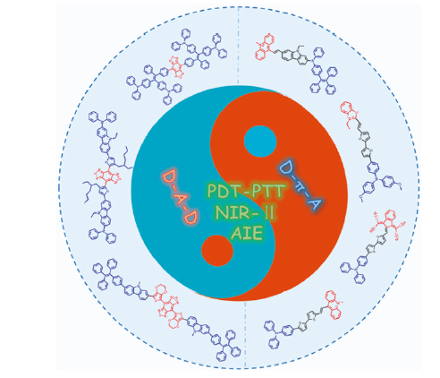
| {{custom_ref.label}} |
{{custom_citation.content}}
{{custom_citation.annotation}}
|
/
| 〈 |
|
〉 |


 AI Summary

