 PDF(13357 KB)
 PDF(13357 KB)
PDF(13357 KB)
PDF(13357 KB)
可激活的NIR-Ⅱ探针用于肿瘤成像
 ({{custom_author.role_cn}}), {{javascript:window.custom_author_cn_index++;}}
Activatable NIR-Ⅱ Probe for Tumor Imaging
({{custom_author.role_en}}), {{javascript:window.custom_author_en_index++;}}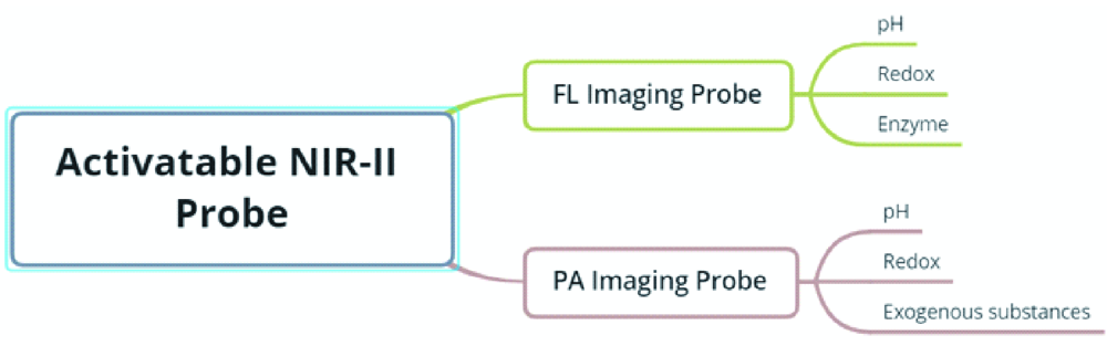
| {{custom_ref.label}} |
{{custom_citation.content}}
{{custom_citation.annotation}}
|
/
| 〈 |
|
〉 |


 AI Summary

