 PDF(14623 KB)
 PDF(14623 KB)
PDF(14623 KB)
PDF(14623 KB)
基于纳米颗粒的化学发光技术在炎症及肿瘤诊疗中的应用
 ({{custom_author.role_cn}}), {{javascript:window.custom_author_cn_index++;}}
Application of Nanoparticles-Based Chemiluminescence in Diagnosis and Treatment of Inflammation and Tumor
({{custom_author.role_en}}), {{javascript:window.custom_author_en_index++;}}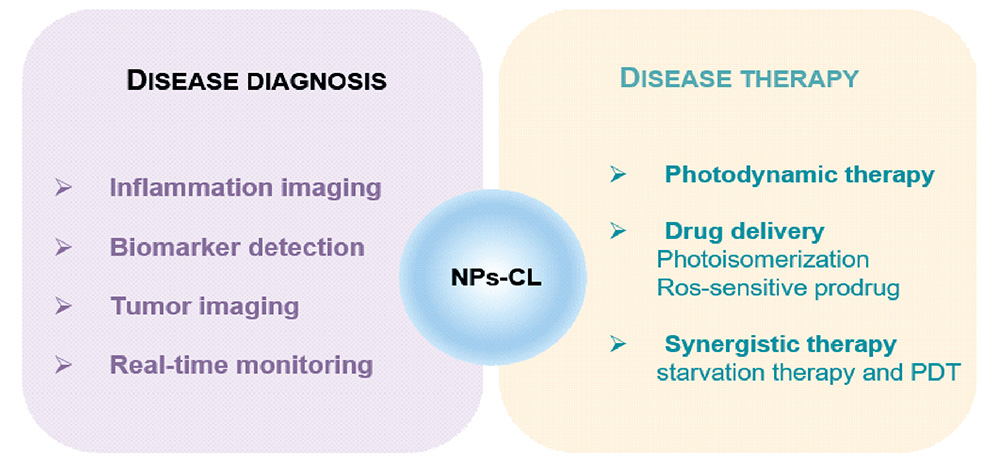
| {{custom_ref.label}} |
{{custom_citation.content}}
{{custom_citation.annotation}}
|
/
| 〈 |
|
〉 |


 AI Summary

