 PDF(11239 KB)
 PDF(11239 KB)
PDF(11239 KB)
PDF(11239 KB)
聚吡咯及其纳米复合材料在光热治疗领域的应用
 ({{custom_author.role_cn}}), {{javascript:window.custom_author_cn_index++;}}
Polypyrrole and Its Nanocomposites Applied in Photothermal Therapy
({{custom_author.role_en}}), {{javascript:window.custom_author_en_index++;}}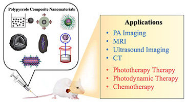
| {{custom_ref.label}} |
{{custom_citation.content}}
{{custom_citation.annotation}}
|
/
| 〈 |
|
〉 |


 AI Summary

