 PDF(3055 KB)
 PDF(3055 KB)
PDF(3055 KB)
PDF(3055 KB)
多功能脂质体递药系统
 ({{custom_author.role_cn}}), {{javascript:window.custom_author_cn_index++;}}
Multifunctional Liposomal Drug Delivery Technology
({{custom_author.role_en}}), {{javascript:window.custom_author_en_index++;}}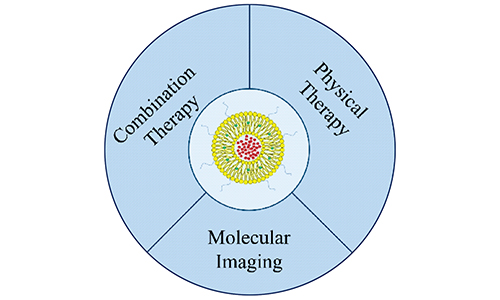
| {{custom_ref.label}} |
{{custom_citation.content}}
{{custom_citation.annotation}}
|
/
| 〈 |
|
〉 |


 AI Summary

