English
新闻公告
More
化学进展 2015, Vol. 27 Issue (10): 1459-1469 DOI: 10.7536/PC150348 前一篇   后一篇

• 综述与评论 •

多光谱光声层析成像及其在生物医学中的应用

刘迎亚, 范霄, 李艳艳, 渠陆陆, 覃海月, 曹英男, 李海涛*   

  1. 江苏师范大学化学化工学院 徐州 221116
  • 收稿日期:2015-03-01 修回日期:2015-05-01 出版日期:2015-10-15 发布日期:2015-09-10
  • 通讯作者: 李海涛 E-mail:haitao@jsnu.edu.cn
  • 基金资助:
    国家自然科学基金项目(No.21375051)和江苏省普通高校研究生科研创新计划项目(No.KYLX_1435)资助

Multispectral Photoacoustic Tomography and Its Development in Biomedical Application

Liu Yingya, Fan Xiao, Li Yanyan, Qu Lulu, Qin Haiyue, Cao Yingnan, Li Haitao*   

  1. School of Chemistry and Chemical Engineering, Jiangsu Normal University, Xuzhou 221116, China
  • Received:2015-03-01 Revised:2015-05-01 Online:2015-10-15 Published:2015-09-10
  • Supported by:
    The work was supported by the National Natural Science Foundation of China (No.21375051) and the Colleges and Universities in Jiangsu Province Plans to Graduate Research and Innovation(No.KYLX_1435).
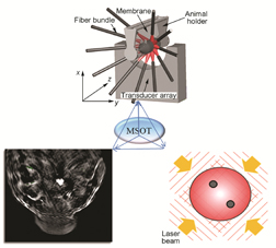
多光谱光声层析成像(MSOT)技术是一种将多光谱成像与光声层析成像(PACT)技术相结合的新技术,该技术利用不同生物组织的光谱吸收特性,用多组不同波长的短脉冲激光照射组织以产生组织特异性的光声信号,从而更好地进行光声成像和组分识别。MSOT兼具光学成像的高灵敏度、高分辨率优势和超声成像可对数厘米深组织成像的长处,同时又能弥补光学成像深度有限和超声成像对比度差的短处,能够实现深层组织的高分辨率、高对比度、高穿透深度的实时无损伤成像。迄今为止,MSOT已应用于肿瘤内光吸收粒子的检测、血管结构和血液氧合作用的评价、生物荧光蛋白的成像以及乳腺癌患者检测的初步研究。随着光声成像系统的不断改进,MSOT与生物标记物(如荧光试剂、金纳米颗粒等)结合对体内分子进行成像,在生物医学中得到了广泛的应用。本文简要综述了MOST的成像原理、实验装置及其性能特点,着重总结了其在生物医学领域的最新应用进展,尤其是在新生血管成像、肿瘤的早期诊断及肿瘤的原位成像方面。
Multispectral photoacoustic tomography is one of the macroscopic observation methods combining multispectral imaging with photoacoustic computed tomography (PACT) technology. Based on the specific spectral absorption characteristics of different biological tissue, the technology illuminates the tissue with multiple sets of short pulse laser of different wavelengths to produce tissue-specific photoacoustic signal for better photoacoustic imaging and component identification. MSOT combines the high sensitivity and resolution of optical imaging with the ability of the several centimeters deep-tissue imaging by ultrasound imaging, while compensated for the depth limitation of optical imaging and the low contrast of ultrasound imaging. Thus it enables noninvasive real-time imaging in the deep tissue with high-resolution and high-contrast, large penetration depths. Over the last years, MSOT has been applied to detecting the light-absorbing particles within the tumor, evaluating vascular structures and blood oxygenation, imaging the biological fluorescent protein and preliminary studies on the breast cancer patients. With the continuous improvement of photoacoustic imaging system, multispectral photoacoustic imaging technology combined with biomarkers (such as fluorescent reagents, gold particles, etc.) has vast applications in biomedical field, especially to molecular imaging in vivo. In this paper, we gives a brief count of the imaging principle, experimental setup and the performance characteristics of MOST. Furthermore, we mainly summarizes the progress of its latest applications in the biomedical field, particularly in the angiogenesis imaging, early diagnosis of cancer and tumors in situ imaging.

Contents
1 Introduction
2 Photoacoustic imaging technology
2.1 Principle of operation
2.2 Classification of photoacoustic imaging
3 Multispectral photoacoustic tomography
3.1 MSOT experimental setup
3.2 Exogenous contrast agents
3.3 Performance characteristics
4 Advantages and challenges of MSOT
5 Applications of MSOT in biomedicine
5.1 Real-time monitoring of neovascularization
5.2 Early diagnosis of cancer
5.3 Targeting tumor imaging
6 Conclusion and outlook

中图分类号: 

()
[1] Bell A G. Am. J. Sci., 1880, 118: 305.
[2] Hu S, Maslov K, Tsytsarev V, Wang L V. J. Biomed. Opt., 2009,14: 040503.
[3] Song L, Maslov K, Wang L V. Opt. Lett., 2010, 35: 1482.
[4] Chen Z, Yang S, Xing D. Opt. Lett., 2012, 37: 3414.
[5] Zhang H F, Maslov K, Wang L V. Nat. Protoc., 2007, 2: 797.
[6] Liao L D, Li M L, Lai H Y, Shih Y Y I, Lo Y C, Tsang S, Chen Y Y. Neuroimage, 2010, 52: 562.
[7] Hu S, Yan P, Maslov K, Lee J M, Wang L V. Opt. Lett., 2009, 34: 899.
[8] Zhang H F, Maslov K, Stoica G, Wang L V. Nat. Biotechnol., 2006, 24: 848.
[9] Zemp R J, Song L, Bitton R, Shung K K, Wang L V. Opt. Express, 2008, 16: 18551.
[10] Zhang J, Yang S, Ji X, Zhou Q, Xing D. Journal of the American College of Cardiology, 2014, 64: 385.
[11] Nie L, Wang S, Wang X, Rong P, Ma Y, Liu G, Huang P, Lu G, Chen X. Small, 2014,10: 1585.
[12] Manohar S, Vaartjes S E, van Hespen J C G, Klaase J M, van den Engh F M, Steenbergen W, Van Leeuwen T G. Opt. Express, 2007, 15: 12277.
[13] Herzog E, Taruttis A, Beziere N, Lutich A A, Razansky D, Ntziachristos V. Radiology, 2012, 263: 461.
[14] Laufer J, Johnson P, Zhang E, Treeby B, Cox B, Pedley B, Beard P.J. Biomed. Opt., 2012, 17: 0560161.
[15] Yang X, Wang L V. J. Biomed. Opt., 2008, 13: 044009.
[16] Scarfe L, Rak-Raszewska A, Geraci S, Darssan D, Sharkey J, Huang J, Burton N C, Mason D, Ranjzad P, Kenny S, Gretz N, Lévy R, Park B K, García-Fiñana M, Woolf A S, Murray P, Wilm B. Sci. Rep., 2015, 5: 1.
[17] Razansky D, Vinegoni C, Ntziachristos V. Phys. Med. Biol., 2009, 54: 2769.
[18] Li P C, Wang C R C, Shieh D B, Wei C W, Liao C K, Poe C, Wu Y N. Opt. Express, 2008, 16: 18605.
[19] Taruttis A, Herzog E., Razansky D, Ntziachristos V. Opt. Express, 2010,17: 21414.
[20] Ntziachristos V, Razansky D. Chem. Rev., 2010, 110: 2783.
[21] 龚小竞(Gong X J), 孟静(Meng J), 白晓淞(Bai X S), 郑加祥(Zheng J X), 宋亮(Song L). 中国医疗器械信息(China Medical Device Information), 2013, 19: 1.
[22] 何军锋(He J F), 谭毅(Tan Y). 激光技术(Laser Technology), 2007, 31: 530.
[23] 谭波(Tan B), 胡建明(Hu J M), 杨盼(Yang P), 丁帅军(Ding S J), 朱仁江(Zhu R J). 激光与光电子学进展(Laser & Optoelectronics Progress), 2013, 50: 39.
[24] Kim C, Favazza C, Wang L V. Chem. Rev., 2010, 110: 2756.
[25] Yao J, Wang L V. Laser Photonics Rev., 2013, 7: 758.
[26] Nie L, Huang P, Li W, Yan X, Jin A, Wang Z, Tang Y X, Wang S J, Zhang X F, Niu G, Chen X Y. ACS Nano, 2014, 8: 12141.
[27] Yoon T J, Cho Y S. World Journal of Gastrointestinal Endoscopy, 2013, 5: 534.
[28] Razansky D, Buehler A, Ntziachristos V. Nat. Protoc., 2011, 6: 1121.
[29] 李莉(Li L), 谢文明(Xie W M), 李晖(Li H). 激光与光电子学进展(Laser & Optoelectronics Progress), 2012, 49: 65.
[30] Oraevsky A A, Jacques S L, Tittel F K. Applied Optics, 1997, 36: 402.
[31] Xia W, Piras D, Heijblom M, van Hespen J C, van Veldhoven S, Prins C, Steenbergen W, van Leeuwen T G, Manohar S. International Society for Optics and Photonics, 2011, 6: 80900L.
[32] Liao W, Liu W, Rogers J E, Usmani F, Tang Y, Wang B, Jiang H, Xie H. Solid-State Sensors, Actuators and Microsystems. 2013 Transducers & Eurosensors XXVII: The 17th International Conference on IEEE, 2013. 1831.
[33] Chee R K, Sampaleanu A, Rishi D, Zemp R J. IEEE Trans. Ultrason. Ferroelectr. Freq., 2014, 61: 1393.
[34] Schwarz M, Buehler A, Ntziachristos V. J. Biophotonics, 2014, 9999.
[35] Dima A, Burton N C, Ntziachristos V. J. Biomed. Opt., 2014, 19: 036021.
[36] Buehler A, Herzog E, Razansky D, Ntziachristos V. Opt. Lett., 2010, 35: 2475.
[37] Taruttis A, Herzog E, Razansky D, Ntziachristos V. Opt. Express, 2010, 18: 19592.
[38] Kruger R A, Liu P, Appledorn C R. Med. Phys., 1995, 22: 1605.
[39] Köstli K P, Frenz M, Bebie H, Weber H P. Phys. Med. Biol., 2001, 46: 1863.
[40] Paltauf G, Viator J A, Prahl S A, Jacques S L. J. Acoust. Soc. Am., 2002, 112: 1536.
[41] Jiang H, Yuan Z, Gu X. JOSA A, 2006, 23: 878.
[42] Rosenthal A, Razansky D, Ntziachristos V. IEEE Trans. Med. Imaging, 2010, 29: 1275.
[43] Paige C C, Saunders M A. ACM Trans. Math. Software, 1982, 8: 43.
[44] Jetzfellner T, Razansky D, Rosenthal A, Schulz R, Englmeier K H, Ntziachristos V. Appl. Phys. Lett., 2009, 95: 013703.
[45] Peng F, Yu D, Luo J. Mech. Syst. Sig. Process., 2011, 25: 549.
[46] Razansky D, Ntziachristos V. Med. Phys., 2007, 34: 4293.
[47] Kruger R A, Kiser W L, Reinecke D R, Kruger G A, Miller K D. Mol. Imag., 2003, 2: 113.
[48] Razansky D, Distel M, Vinegoni C, Ma R, Perrimon N, Köster R W, Ntziachristos V. Nat. Photonics, 2009, 3: 412.
[49] Rosenthal A, Razansky D, Ntziachristos V. IEEE Trans. Med. Imaging, 2009, 28: 1997.
[50] Taruttis A, Rosenthal A, Kacprowicz M, Burton N C, Ntziachristos, V. IEEE Trans. Med. Imaging, 2014, 33: 1194.
[51] Taruttis A, Claussen J, Razansky D, Ntziachristos V. J. Biomed. Opt., 2012, 17: 0160091.
[52] Weissleder R, Pittet M J. Nature, 2008, 452: 580.
[53] Giepmans B N G, Adams S R, Ellisman M H, Tsien R Y. Science, 2006, 312: 217.
[54] Beard P. Interface focus, 2011,1: 602.
[55] Dima A, Ntziachristos V. Expert Opinion on Medical Diagnostics, 2011, 5: 263.
[56] Qin H, Zhou T, Yang S, Chen Q, Xing D. Nanomed., 2013, 8: 1611.
[57] Razansky D, Vinegoni C, Ntziachristos V. Opt. Lett., 2007, 32: 2891.
[58] Ntziachristos V, Razansky D. Modern Biopharmaceuticals: Recent Success Stories, 2013: 211.
[59] Zhang H F, Maslov K, Stoica G, Wang L V. Nat. Biotechnol., 2006, 24: 848.
[60] Ma R, Taruttis A, Ntziachristos V, Razansky D. Opt. Express, 2009, 17: 21414.
[61] Ermilov S A, Khamapirad T, Conjusteau A, Leonard M H, Lacewell R, Mehta K, Oraevsky A A. J. Biomed. Opt., 2009, 14: 024007.
[62] Niederhauser J J, Jaeger M, Lemor R, Weber P, Frenz M. IEEE Trans. Med. Imaging, 2005, 24: 436.
[63] Taruttis A, Ntziachristos V. Nat. Photonics, 2015, 9: 219.
[64] Li M L, Oh J T, Xie X, Ku G, Wang W, Li, C, Wang L V. Proc. IEEE, 2008, 96: 481.
[65] Ntziachristos V. Nat. Methods, 2010, 7: 603.
[66] Razansky D, Vinegoni C, Ntziachristos V. Proc. SPIE. 2009, 71770D.
[67] Cox B, Laufer J G, Arridge S R, Beard P C. J. Biomed. Opt., 2012, 17: 0612021.
[68] Folkman J. J. Natl. Cancer. Inst., 1990, 82: 4.
[69] Kruger R A, Kiser W L, Reinecke D R, Kruger G A. Med. Phys., 2003, 30: 856.
[70] Wang X, Stoica G, Xie X, Ku G, Wang L V. J. Biomed. Opt., 2006,11: 024015.
[71] Wang X, Pang Y, Ku G, Xie X, Stoica G, Wang L V. Nat. Biotechnol., 2003, 21: 803.
[72] Siphanto R I, Thumma K K, Kolkman R G M, van Leeuwen T G, de Mul F F M, van Neck J W, Steenbergen W. Opt. Express, 2005, 13: 89.
[73] Ku G, Wang X, Xie X, Stoica G, Wang L V. Appl. Opt., 2005, 44:770.
[74] Dima A, Ntziachristos V. Optics express, 2012, 20: 25044.
[75] Razansky D, Harlaar N J, Hillebrands J L, Taruttis A, Herzog E, Zeebregts C J, Ntziachristos V. Mol. Imag. Biol., 2012, 14: 277.
[76] Gottschalk S, Fehm T F, Deán-Ben X L, Razansky D. Journal of Cerebral Blood Flow & Metabolism, 2015, 35: 531
[77] Buehler A, Kacprowicz M, Taruttis A, Ntziachristos V. Opt. Lett., 2013, 38: 1404.
[78] Buehler A, Herzog E, Ale A, Smith B D, Ntziachristos V, Razansky D. EJNMMI Res., 2012, 2: 14.
[79] Taruttis A, Morscher S, Burton N C, Razansky D, Ntziachristos V. PloS One, 2012, 7: e30491.
[80] Morscher S, Burton N C, Taruttis A, Deliolanis N C, Razansky D, Ntziachristos V. SPIE BiOS. 2012,822330.
[81] Burton N C, Patel M, Morscher S, Driessen W H, Claussen J, Beziere N, Ntziachristos V. Neuroimage, 2013, 65: 522.
[82] Huang G, Si Z, Yang S, Li C, Xing D. Mater. Chem., 2012, 22: 22575.
[83] Rayavarapu R G, Petersen W, Ungureanu C, Post J N, van Leeuwen T G, Manohar S. J. Biomed. Imaging, 2007, 2007: 5.
[84] Eghtedari M, Oraevsky A, Copland J A, Kotov N A, Conjusteau A, Motamedi M. Nano Lett., 2007, 7: 1914
[85] De La Zerda A, Zavaleta C, Keren S, Vaithilingam S, Bodapati S, Liu Z, Gambhir S S. Nat. Nanotechnol., 2008, 3: 557.
[86] Kang N Y, Park S J, Ang X W E, Samanta A, Driessen W H, Ntziachristos V, Chang Y T. Chem. Commun., 2014, 50: 6589.
[87] Deliolanis N C, Ale A, Morscher S, Burton N C, Schaefer K, Radrich K, Ntziachristos V. Mol. Imag. Biol., 2014: 1.
[88] Beziere N, Lozano N, Nunes A, Salichs J, Queiros D, Kostarelos K, Ntziachristos V. Biomaterials, 2015, 37: 415.
[89] Chamberland D L, Agarwal A, Kotov N, Fowlkes J B, Carson P L, Wang X. Nanotechnology, 2008, 19: 095101.
[90] Li L, Zemp R J, Lungu G, Wang L V. J. Biomed. Opt., 2007, 12: 020504.
[91] Shashkov E V, Everts M, Galanzha E I, Zharov V P. Nano Lett., 2008, 8: 3953.
[92] Galanzha E I, Shashkov E V, Kelly T, Kim J W, Yang L, Zharov V P. Nat. Nanotechnol., 2009, 4: 855.
[1] 钱雪丹, 余伟江, 付濬哲, 王幽香, 计剑. 透明质酸基微纳米凝胶的制备及生物医学应用[J]. 化学进展, 2023, 35(4): 519-525.
[2] 马佳慧, 袁伟, 刘思敏, 赵智勇. 小分子共价DNA的组装及生物医学应用[J]. 化学进展, 2022, 34(4): 837-845.
[3] 蔡雪儿, 简美玲, 周少红, 王泽峰, 王柯敏, 刘剑波. 人造细胞的化学构建及其生物医学应用研究[J]. 化学进展, 2022, 34(11): 2462-2475.
[4] 王振, 李曦, 栗园园, 王其, 卢晓梅, 范曲立. 可激活的NIR-Ⅱ探针用于肿瘤成像[J]. 化学进展, 2022, 34(1): 198-206.
[5] 任飞, 石建兵, 佟斌, 蔡政旭, 董宇平. 具有聚集诱导发光性质的近红外荧光染料[J]. 化学进展, 2021, 33(3): 341-354.
[6] 赵平平, 杨军星, 施健辉, 朱静怡. 基于树状大分子的SPECT成像造影剂的构建及其应用[J]. 化学进展, 2021, 33(3): 394-405.
[7] 刘加伟, 王婧, 王其, 范曲立, 黄维. 激活型有机光声造影剂的应用[J]. 化学进展, 2021, 33(2): 216-231.
[8] 胡强强, 郭和泽, 窦红静. ZIF-8纳米颗粒的粒径调控及生物医学应用[J]. 化学进展, 2020, 32(5): 656-664.
[9] 肖肖, 陈昌盛, 刘伟强, 张业顺. 丝胶蛋白的结构、性能及生物医学应用[J]. 化学进展, 2017, 29(5): 513-523.
[10] 卢晓梅, 陈鹏飞, 胡文博, 唐玉富, 黄维, 范曲立. 有机光电材料在光声成像领域的应用[J]. 化学进展, 2017, 29(1): 119-126.
[11] 杜凯, 朱艳红, 徐辉碧, 杨祥良. 多功能磁性纳米粒的合成、修饰及生物医学应用[J]. 化学进展, 2011, 23(11): 2287-2298.
[12] 黄毅,黄金花,谢青季,姚守拙. 糖-蛋白质相互作用*[J]. 化学进展, 2008, 20(06): 942-950.