English
新闻公告
More
化学进展 2021, Vol. 33 Issue (3): 341-354 DOI: 10.7536/PC200614 前一篇   后一篇

• 综述 •

具有聚集诱导发光性质的近红外荧光染料

任飞1, 石建兵1,*(), 佟斌1, 蔡政旭1, 董宇平1,*()   

  1. 1 北京理工大学材料学院 北京 100081
  • 收稿日期:2020-06-05 修回日期:2020-06-25 出版日期:2021-03-20 发布日期:2020-11-30
  • 通讯作者: 石建兵, 董宇平
  • 作者简介:
    * Corresponding author e-mail: (Jianbing Shi); (Yuping Dong)
  • 基金资助:
    国家自然科学基金项目(21875019); 国家自然科学基金项目(51673024); 国家自然科学基金项目(21975020); 国家自然科学基金项目(51803009)

Near Infrared Fluorescent Dyes with Aggregation-Induced Emission

Fei Ren1, Jianbing Shi1,*(), Bin Tong1, Zhengxu Cai1, Yuping Dong1,*()   

  1. 1 School of Materials Science and Engineering, Beijing Institute of Technology,Beijing 100081, China
  • Received:2020-06-05 Revised:2020-06-25 Online:2021-03-20 Published:2020-11-30
  • Contact: Jianbing Shi, Yuping Dong
  • Supported by:
    the National Natural Science Foundation of China(21875019); the National Natural Science Foundation of China(51673024); the National Natural Science Foundation of China(21975020); the National Natural Science Foundation of China(51803009)

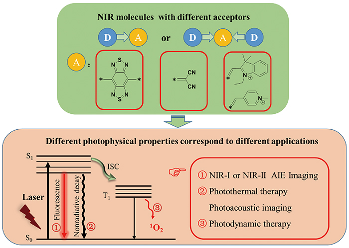
聚集诱导发光(AIE)现象的发现为解决传统有机荧光分子在高浓度和聚集形态下存在的荧光猝灭问题提供了最佳方案,并实现了在光电器件、化学传感、生物成像和靶向治疗等众多领域的广泛应用。随着对AIE发光机理研究的不断深入,AIE分子体系得到了极大的扩展。其中,一类具有给体-受体结构的AIE分子能够显著降低分子能隙,使发光分子波长从可见光区(400~700 nm)延伸到近红外(NIR)区(700~1700 nm)。由于NIR发光分子在生物医学领域中的独特优势,其已成为目前AIE研究的热点。随着对NIR分子设计及应用的不断探索,附加不同功能且发光波长更长的AIE分子也被开发出来了,并实现了对生物体特定组织的NIR荧光成像、光声成像、光动力治疗和光热治疗等。本文总结了近年来具有AIE性能的NIR荧光分子的结构及其在生物医学领域的相关应用。

The discovery of the aggregation-induced emission(AIE) phenomenon provides the best solution to solve the problem of fluorescence quenching of traditional organic fluorescent molecules at high concentrations and aggregation state. AIE molecules are widely used in many fields such as photoelectric devices, chemical sensing, biological imaging and targeting therapy. With the deepening of the research on the emissive mechanism of AIE, the AIE molecular system has been greatly expanded. Among them, a class of AIE molecules with donor-acceptor structures can significantly reduce the molecular energy gap and extend the emission wavelengths of molecules from the visible light region(400~700 nm) to the near infrared(NIR) region(700~1700 nm). Due to the unique advantages of NIR fluorescent molecules in the field of biomedicine, they have become the hot topic of AIE research. With the continuous exploration of the design and application of NIR molecules, AIE molecules with different functions and longer emission wavelengths have also been developed, and realized the application of NIR fluorescence imaging, photoacoustic imaging, photodynamic therapy and photothermal therapy to specific tissues of organisms. This article summarizes the structure of NIR fluorescent molecules with AIE performance in recent years and their related applications in the field of biomedicine.

Contents

1 Introduction

2 The discovery and mechanism of aggregation-induced emission

3 The advantages of NIR and the partition of fluorescent windows

4 Principle of molecular design of NIR dyes

5 NIR fluorescent dyes with AIE property and their applications

5.1 Design and application of benzothiadiazole NIR dyes

5.2 Design and application of malononitrile NIR dyes

5.3 Design and application of ionic NIR dyes

6 Conclusion

()
图1 (a)不同波段的光的组织穿透深度;(b)进入组织时,不同发光波长的荧光团对组织的穿透深度及被生物组织反射和吸收情况[12]
Fig.1 (a) Tissue penetration depth of light with different wavelengths;(b) light when entering a tissue can be reflected or absorbed by molecules within the tissue or excite fluorophores to emit light at different wavelengths[12]. Copyright 2018, Royal Society of Chemistry
图2 (a)TB1的化学结构;(b)TB1和IR-26在溶液态和粉末状态下的NIR-Ⅱ荧光图像(溶液:DMSO);(c)TB1在THF中的紫外吸收光谱和荧光发射光谱;(d)TB1在不同水含量的THF/水混合物中的荧光强度比;(e)TB1-RGD纳米粒子的制备路线;(f)对完整头皮和头骨的小鼠进行大脑无创NIR-Ⅱ荧光成像(808 nm, 60 mW·cm-2)[35]
Fig.2 (a) Chemical structure of TB1;(b) NIR-Ⅱ fluorescence images of TB1 and IR-26 in solution and powder states(solution: DMSO);(c) UV absorption and fluorescence spectra of TB1 in THF solution;(d) fluorescence intensity ratio of TB1 in THF/water mixture with different water contents;(e) preparation route of TB1-RGD nanoparticle;(f) brain non-invasive NIR-Ⅱ fluorescence imaging on mice with intact scalp and skull(808 nm, 60 mW·cm-2)[35]. Copyright 2018, Wiley
图3 (a)HLZ-BTED的合成路线;(b)HLZ-BTED在不同水含量(0%至95%)的荧光发射光谱(Ex: 785 nm);(c)尾静脉注射HLZ-BTED纳米粒子2 min后,获得了4T1荷瘤小鼠的NIR-Ⅱ肿瘤血管荧光图像[36]
Fig.3 (a) Synthesis route of HLZ-BTED;(b) fluorescence emission spectra of HLZ-BTED at different water contents(0% to 95%)(Ex: 785 nm);(c) the NIR-Ⅱ tumor blood vessel fluorescence images of 4T1 tumor-bearing mice 2 minutes after injection of HLZ-BTED nanoparticles into the tail vein[36]. Copyright 2019, Royal Society of Chemistry
图4 (a)HQL1和HQL2的化学结构;(b)DCM中HQL1和HQL2的UV-vis-NIR吸收光谱和NIR-Ⅱ荧光光谱;(c)U87MG荷瘤裸鼠的荧光成像(808 nm,90 mW·cm-2);(d)HQL2纳米粒子成像的毛细血管的横截面强度(黑线)和高斯拟合荧光强度分布图(红线)(c中的红色虚线)[37]
Fig.4 (a) The chemical structure of HQL1 and HQL2;(b) UV-vis-NIR absorption spectra and NIR-Ⅱ fluorescence spectra of HQL1 and HQL2 in DCM;(c) fluorescence imaging of U87MG tumor-bearing nude mice(808 nm, 90 mW·cm-2);(d) cross-sectional intensity of capillaries imaged by HQL2 nanoparticles(black line) and Gaussian fitted fluorescence intensity distribution map(red line)(red dotted line in c)[37].Copyright 2020, Royal Society of Chemistry
图5 (a)HL1-HL3的化学结构;(b~d)尾静脉注射HL3纳米粒子(200 μL,1.5 mg·mL -1)后,在不同LP滤光片和曝光时间下C57BL/6小鼠中的脑血管的NIR-Ⅱ和NIR-Ⅱb荧光图像:(b)1000 nm LP,4 ms,90 mW·cm-2;(c)1250 nm LP,60 ms,90 mW·cm-2;(d)1550 nm LP,500 ms,90 mW·cm-2;(e~g)毛细血管的横截面强度(红线)和高斯拟合荧光强度分布图(黑线)[38]
Fig.5 (a) The chemical structure of the target molecule;(b~d) NIR-Ⅱ and NIR-Ⅱb fluorescence images of cerebral vasculature with different LP filters in C57BL/6 mice(n = 3) after tail intravenous injection of HL3 dots(200 μL, 1.5 mg·mL -1):(b) 1000 nm LP, 4 ms exposure time, and 90 mW·cm-2;(c) 1250 nm LP, 60 ms exposure time, and 90 mW·cm-2;(d) 1550 nm LP, 500 ms exposure time, and 90 mW·cm-2;(e~g) the fluorescence intensity profiles fitted using Gaussian, cross-section intensity(black lines), and the tiny vessel(red-dashed lines)[38]. Copyright 2020, Royal Society of Chemistry
图6 (a)目标分子2TT-oC6B的化学结构及制备AIE纳米粒子的原理图;(b)去离子水中2TT-oC6B纳米粒子的紫外吸收和荧光发射图谱;(c)在兔子模型的输尿管内注射2TT-oC6B纳米粒子实现对输尿管的NIR-Ⅱ荧光成像(808 nm激发);(d)2TT-oC6B纳米粒子(红色虚线)和ICG(蓝色虚线)对输尿管成像质量的比较[39]
Fig.6 (a) The chemical structure of the target molecule 2TT-oC6B and the schematic diagram of the preparation of AIE nanoparticles;(b) UV absorption and fluorescence emission spectra of 2TT-oC6B nanoparticles in deionized water;(c) intraureteral injection of 2TT-oC6B nanoparticles to achieve NIR-Ⅱ fluorescence imaging of the ureter in the rabbit model(Ex: 808 nm);(d) comparison of 2TT-oC6B nanoparticles(red dotted line) and ICG(blue dotted line) on ureter imaging quality[39]. Copyright 2020, American Chemical Society
图7 (a)SYL分子的化学结构及NIR-Ⅱ在可见光和紫外灯下的荧光图像;(b)对4T1荷瘤小鼠尾静脉注射SYL纳米粒子后0、2、8、12和24 h的体内NIR-Ⅱ荧光和PA图像(808 nm,82 mW·cm-2)[40]
Fig.7 (a) Chemical structure of SYL molecule and NIR-Ⅱ fluorescence pictures in visible light and ultraviolet light;(b) after injection of SYL nanoparticles into the tail vein of 4T1 tumor-bearing mice for 0, 2, 8, 12 and 24 h, in vivo NIR-Ⅱ fluorescence and PA images(808 nm, 82 mW·cm-2)[40]. Copyright 2019, Royal Society of Chemistry
图8 光敏剂TQ-BTPE的化学结构和NIR-Ⅱ光激活的双光子光动力癌细胞消融的示意图[41]
Fig.8 Chemical structure of photosensitizer TQ-BTPE and schematic illustration of NIR-Ⅱ light activated two-photon photodynamic cancer cell ablation[41]. Copyright 2020, Wiley
图9 (a)目标分子结构式及QM-1、QM-2、QM-3的单晶结构;(b)静脉注射QM-5(0.15 mg/kg)后不同时间段的荷瘤小鼠的体内无创成像;(c)尾静脉注射QM-5(0.15 mg/kg)24 h后,荷瘤小鼠的3D荧光成像[48]
Fig.9 (a) Target molecular structures and single crystal structures of QM-1, QM-2, and QM-3;(b) in vivo non-invasive imaging of tumor-bearing mice at different time after intravenous injection of QM-5(0.15 mg/kg);(c) 3D fluorescence imaging of tumor-bearing mice after the tail vein injection of QM-5(0.15 mg/kg) for 24 hours[48]. Copyright 2015, Wiley
图10 设计用于淀粉样蛋白-β斑块检测的AIE探针。 (a)基于always-on模式的商业探针ThT;(b,c)解决商业探针ThT固有缺陷并创建超灵敏的off-on NIR探针的“分步”策略:(i)引入亲脂性π共轭噻吩桥以将波长扩展到NIR区域并且具有血脑屏障渗透性;(ii)将ACQ替换为AIE结构单元,以及(iii)调整磺酸酯取代的位置,以确保在与淀粉样蛋白-β斑块结合之前保持荧光关闭状态;(d~g)分别使用ThS和QM-FN-SO3对野生小鼠和阿尔茨海默病(AD)模型(APP/PS1转基因)小鼠海马区的脑切片进行组织学染色;(h)穿过大脑切片线性区域(ROI)的强度分布;(i)DCM-N、QM-FN和QM-FN-SO3的信噪比[49]
Fig.10 Rational design of NIR AIE-active probes for Aβ deposition. (a) Commercial probe ThT based on thealways-on pattern;(b,c) the“step-by-step” strategy to address the inherent defects of commercial ThT and create ultrasensitive off-on NIR probes:(i) introducing lipophilic π-conjugated thiophene-bridge for extending the wavelength to the NIR region with BBB penetrability,(ii) replacing the ACQ to AIE building block, and(iii) tuning the sulfonate substituted position for guaranteeing fluorescence-off state before binding to Aβ deposition.;(d~g) histological staining of the brain slices in the hippocampus region from wild-type mice and Alzheimer’s disease(AD)-model(APP/PS1 transgenic) mice using ThS and QM-FN-SO3, respectively;(h) the intensity profiles of the linear regions of interest(ROI) crossing the brain slices;(i) the S/N ratios of DCM-N, QM-FN, and QM-FN-SO3[49]. Copyright 2019, American Chemical Society
图11 (a)分子TFM的化学合成路线;(b)通过纳米沉积法制备TFM纳米粒子;(c)TFM纳米粒子用于光声成像指导癌症的PTT-PDT示意图[50]
Fig.11 (a) Chemical synthesis routes of TFM;(b) preparation of TFM nanoparticles by nanodeposition method;(c) illustration of the use of TFM NPs for PAI-guided PTT-PDT cancer theranostics[50]. Copyright 2019, Wiley
图12 (a)目标分子的化学合成路线;(b)AGL AIE dots的NIR余辉发光机理的示意图;(c)NIR余辉成像引导癌症手术示意图[51]
Fig.12 (a) Synthesis routes of the target molecules;(b) schematic diagram of the NIR afterglow luminescence mechanism of AGL AIE dots;(c) schematic diagram of cancer surgery guided by NIR afterglow imaging[51]. Copyright 2019, American Chemical Society
图13 设计用于细菌跟踪和PDT的TPACN-D-Ala探针;(a)D-Ala与TPACN发生化学反应制备TPACN-D-Ala探针;将其静脉注射到感染细菌的小鼠中,以实现特定的荧光成像和图像引导抗菌;(b)通过血液循环到达受感染的组织部位,TPACN-D-Ala被整合到肽聚糖中以产生强烈的NIR荧光;此外,可以在体内实现侵入性细菌的特异性成像和有效治疗;(c)PBS中TPACN-D-Ala和TPACN的紫外线吸收和荧光发射图谱;(d)以ABDA为参考测试TPACN-D-Ala(10 μM)和Ce6(10 μM)ROS的产生(白光,60 mW·cm -2,A0和A是分子在378 nm处的吸光度)[52]
Fig.13 Design and characterization of the developed TPACN-D-Ala probe for bacterial tracking and photodynamic therapy(PDT);(a) TPACN-D-Ala was synthesized by combining D-Ala with AIE photosensitizer TPACN, which could be intravenously injected into the bacteria-infected mouse to realize specific fluorescence light-up and image-guided antibacterial PDT;(b) Once reaching the infected tissue site through blood circulation, TPACN-D-Ala would be integrated into peptidoglycan to produce intense NIR fluorescence, specific imaging and efficient treatment of invasive bacteria could be achieved in vivo;(c) UV absorption and fluorescence emission of TPACN-D-Ala or TPACN in PBS;(d) measurement of 1O2 production of TPACN-D-Ala(10 μM) or Ce6(10 μM) using ADBA under light irradiation(white light, 60 mW·cm -2). ABDA(black) solutions were used as control; A0 and A are the absorbance of ABDA at 378 nm[52]. Copyright 2020, Royal Society of Chemistry
图14 UCNP@TTD cRGD NPs的制备;在近红外激光照射下体外3D癌细胞球体和小鼠肿瘤模型中深部肿瘤的NIR荧光成像和PDT的应用示意图[53]
Fig.14 Preparation of UCNP@TTD cRGD NPs; schematic diagram of NIR fluorescence imaging and PDT application of deep tumors in 3D cancer cell spheres and mouse tumor models in vitro under NIR laser irradiation[53]. Copyright 2019, Ivyspring
图15 (a)目标分子的化学结构式;(b)在相同条件下将TPP-1、TPP-2和TPP-3与HeLa共培养后成像的实时视频截图[TPP-1] = [TPP-2] = [TPP-3] = 1.0×10⁻⁷ mol/L[54]
Fig.15 (a) The chemical structures of the target molecules;(b) real-time video screenshot of TPP-1, TPP-2 and TPP-3 co-cultured with HeLa under the same conditions [TPP-1] = [TPP-2] = [TPP-3] = 1.0×10⁻⁷ mol/L[54]. Copyright 2018, Royal Society of Chemistry
图16 (a)PMTi合成的示意图;(b)线粒体靶向的亚细胞药物递送[55]
Fig.16 (a) Schematic diagram of PMTi synthesis;(b) mitochondrial-targeted subcellular drug delivery[55]. Copyright 2019, Wiley
图17 (a)TPE-DPA-TCyP的化学结构;(b)TPE-DPA-TCyP作为抗肿瘤免疫ICD诱导剂的作用机制[56]
Fig.17 (a) The chemical structure of TPE-DPA-TCyP;(b) the proposed mechanism of TPE-DPA-TCyP as an effective ICD inducer for antitumor immunity[56]. Copyright 2019, Wiley
图18 (a)目标分子的化学结构式;(b)不同溶剂配比的相对发射强度(I/I0)图谱,插图:在365 nm紫外线照射下,在DMSO溶液和DMSO/甲苯混合物(含95%甲苯)中的TTPy荧光照片;(c)不同组在治疗后不同时间点的肿瘤体积生长曲线;(d)测量每组中的小鼠体重随时间的变化[57]
Fig.18 (a) Chemical structures of target molecules;(b) relative emission intensity(I/I0) spectra of different solvent ratios, inset: fluorescence photos of TTPy in DMSO solution and DMSO/toluene mixture(containing 95% toluene) under UV irradiation at 365 nm;(c) the volume growth curves of tumors at different time points post-treatment in different groups;(d) body weight measurement of the mice in each group[57]. Copyright 2018, Wiley
图19 (a)分子TFPy、TFVP和TPE-TFPy的化学结构式;(b)使用三种AIE分子实现“1+1+1>3”协同增强光动力疗法的示意图[58]
Fig.19 (a) Chemical structures of TFPy, TFVP and TPE-TFPy;(b) schematic illustration of using three AIEgens for achieving “1+1+1>3” synergistic enhanced photodynamic therapy [58]. Copyright 2020, Wiley
图20 (a)目标分子的化学结构式;(b,c)在含有100 μM Hg 2+的PBS中培养40 min或(d,e)仅在PBS中培养40 minAS2CP-TPA对HeLa细胞染色的共聚焦图像,培养之前(伪红色)和培养之后(伪绿色)(λex=488 nm,λem=600~750 nm)[59]
Fig.20 (a) The chemical structure of the target molecule; overlaid confocal images of AS2CP-TPA-stained HeLa cells before(pseudo red) and after(pseudo green) incubation in PBS containing 100 μM Hg 2+ for 40 min(b, c) or incubation in PBS only for 40 min(d, e)(λex = 488 nm and λ em= 600~750 nm)[59]. Copyright 2018, Royal Society of Chemistry
图21 使用TTVP进行革兰氏阳性细菌的超快速判别和高效光动力抗菌疗法的示意图[60]
Fig.21 Schematic illustration of using AIEgen TTVP for ultrafast discrimination of Gram-positive bacteria and highly efficient photodynamic antibacterial therapy[60]. Copyright 2020, Elsevier
[1]
Luo J D, Xie Z L, Lam J W Y, Cheng L, Tang B Z, Chen H Y, Qiu C F, Kwok H S, Zhan X W, Liu Y Q, Zhu D B. Chem. Commun., 2001, (18):1740.
[2]
Fan X, Sun J L, Wang F Z, Chu Z Z, Wang P, Dong Y Q, Hu R R, Tang B Z, Zou D C. Chem. Commun., 2008, (26):2989.
[3]
Feng H T, Yuan Y X, Xiong J B, Zheng Y S, Tang B Z. Chem. Soc. Rev., 2018, 47(19):7452.
[4]
Zhang Y H, Mao H L, Xu W Q, Shi J B, Cai Z X, Tong B, Dong Y P. Chem. Eur. J., 2018, 24(60):15965.
[5]
Feng X, Tong B, Shen J B, Shi J B, Han T, Chen L, Zhi J G, Lu P, Ma Y G, Dong Y P. J. Phys. Chem. B, 2010, 114(50):16731.
[6]
He J T, Xu B, Chen F P, Xia H J, Li K P, Ye L, Tian W J. J. Phys. Chem. C, 2009, 113(22):9892.
[7]
Mei J, Hong Y N, Lam J W Y, Qin A J, Tang Y H, Tang B Z. Adv. Mater., 2014, 26(31):5429.
[8]
Hu R R, Leung N L C, Tang B Z. Chem. Soc. Rev., 2014, 43(13):4494.
[9]
Wang D, Tang B Z. Acc. Chem. Res., 2019, 52:2559.
[10]
Guo Z Q, Park S, Yoon J, Shin I. Chem. Soc. Rev., 2014, 43(1):16.
[11]
Gao M, Yu F B, Lv C, Choo J, Chen L X. Chem. Soc. Rev., 2017, 46(8):2237.
[12]
He S Q, Song J, Qu J L, Cheng Z. Chem. Soc. Rev., 2018, 47(12):4258.
[13]
Hong G S, Antaris A L, Dai H J. Nat. Biomed. Eng., 2017, 1:0010.
[14]
Wang X D, Wang S M, Lv J, Shao S Y, Wang L X, Jing X B, Wang F S. Chem. Sci., 2019, 10(10):2915.
[15]
Huang G X, Xia Q, Huang W B, Tian J W, He Z K, Li B S, Tang B Z. Angew. Chem. Int. Ed., 2019, 58(49):17814.
[16]
Guo X M, Yuan P S, Qiao X F, Yang D Z, Dai Y F, Sun Q, Qin A J, Tang B Z, Ma D G. Adv. Funct. Mater., 2020, 30(9):1908704.
[17]
Wei G Q, Tao Y C, Wu J J, Li Z Z, Zhuo M P, Wang X D, Liao L S. J. Phys. Chem. Lett., 2019, 10:679.
[18]
Shahbazi M A, Faghfouri L, Ferreira M P A, Figueiredo P, Maleki H, Sefat F, Hirvonen J, Santos H A. Chem. Soc. Rev., 2020, 49(4):1253.
[19]
Cheng H B, Li Y Y, Tang B Z, Yoon J. Chem. Soc. Rev., 2020, 49(1):21.
[20]
Mei J, Leung N L C, Kwok R T K, Lam J W Y, Tang B Z. Chem. Rev., 2015, 115(21):11718.
[21]
Hong Y N, Lam J W Y, Tang B Z. Chem. Commun., 2009(29):4332.
[22]
Hong Y N, Lam J W Y, Tang B Z. Chem. Soc. Rev., 2011, 40(11):5361.
[23]
Ding D, Li K, Liu B, Tang B Z. Acc. Chem. Res., 2013, 46(11):2441.
[24]
Chi Z G, Zhang X Q, Xu B J, Zhou X, Ma C P, Zhang Y, Liu S W, Xu J R. Chem. Soc. Rev., 2012, 41(10):3878.
[25]
Tu Y J, Liu J K, Zhang H K, Peng Q, Lam J W Y, Tang B Z. Angew. Chem. Int. Ed., 2019, 58(42):14911.
[26]
Tian R, Ma H, Yang Q, Wan H, Zhu S, Chandra S, Sun H, Kiesewetter D O, Niu G, Liang Y, Chen X. Chem. Sci. 2019, 10:326.
[27]
Zhang X, An L, Tian Q W, Lin J M, Yang S P. J. Mater. Chem. B, 2020, 8(22):4738.
[28]
Staderini M, Martín M A, Bolognesi M L, MenÉndez J C. Chem. Soc. Rev., 2015, 44(7):1807.
[29]
Kaur M, Choi D H. Chem. Soc. Rev., 2015, 44(1):58.
[30]
Sun W, Guo S G, Hu C, Fan J L, Peng X J. Chem. Rev., 2016, 116(14):7768.
[31]
Kowada T, Maeda H, Kikuchi K. Chem. Soc. Rev., 2015, 44(14):4953.
[32]
Toutchkine A, Nguyen D V, Hahn K M. Org. Lett., 2007, 9(15):2775.
[33]
Samanta A, Vendrell M, Das R, Chang Y T. Chem. Commun., 2010, 46(39):7406.
[34]
Ding F, Zhan Y B, Lu X J, Sun Y. Chem. Sci., 2018, 9(19):4370.
[35]
Sheng Z H, Guo B, Hu D H, Xu S D, Wu W B, Liew W H, Yao K, Jiang J Y, Liu C B, Zheng H R, Liu B. Adv. Mater., 2018, 30(29):1870214.
[36]
Lin J C, Zeng X D, Xiao Y L, Tang L, Nong J X, Liu Y F, Zhou H, Ding B B, Xu F C, Tong H X, Deng Z X, Hong X C. Chem. Sci., 2019, 10(4):1219.
[37]
Li Q Q, Ding Q H, Li Y, Zeng X D, Liu Y S, Lu S Y, Zhou H, Wang X F, Wu J Z, Meng X L, Deng Z X, Xiao Y L. Chem. Commun., 2020, 56(22):3289.
[38]
Li Y, Liu Y F, Li Q Q, Zeng X D, Tian T, Zhou W Y, Cui Y, Wang X K, Cheng X D, Ding Q H, Wang X F, Wu J Z, Deng H, Li Y Q, Meng X L, Deng Z X, Hong X C, Xiao Y L. Chem. Sci., 2020, 11(10):2621.
[39]
Du J, Liu S J, Zhang P F, Liu H X, Li Y Y, He W, Li C B, Chau J H C, Kwok R T K, Lam J W Y, Cai L T, Huang Y H, Zhang W J, Hou J Q, Tang B Z. ACS Appl. Mater. Interfaces, 2020, 12(7):8040.
[40]
Zhang R P, Xu Y L, Zhang Y, Kim H S, Sharma A, Gao J, Yang G F, Kim J S, Sun Y. Chem. Sci., 2019, 10(36):8348.
[41]
Wang S W, Chen H, Liu J, Chen C J, Liu B. Adv. Funct. Mater., 2020, 30(30):2002546.
[42]
Redon S, Eucat G, Ipuy M, Jeanneau E, Gautier-Luneau I, Ibanez A, Andraud C, Bretonnière Y. Dye. Pigment., 2018, 156:116.
[43]
Ren F, Shi J B, Tong B, Cai Z X, Dong Y P. Mater. Chem. Front., 2019, 3(10):2072.
[44]
Li X J, Yao J, Angunawela I, Sun C K, Xue L W, Liebman-Pelaez A, Zhu C H, Yang C H, Zhang Z G, Ade H, Li Y F. Adv. Energy Mater., 2018, 8(23):1800815.
[45]
Yang Y K, Qiu B B, Chen S S, Zhou Q J, Peng Y, Zhang Z G, Yao J, Luo Z H, Chen X F, Xue L W, Feng L L, Yang C, Li Y F. J. Mater. Chem. A, 2018, 6(20):9613.
[46]
Guo Z Q, Shao A D, Zhu W H. J. Mater. Chem. C, 2016, 4(14):2640.
[47]
Lei Y X, Lai Y Y, Dong L C, Shang G J, Cai Z X, Shi J B, Zhi J G, Li P F, Huang X B, Tong B, Dong Y P. Chem. Eur. J., 2018, 24(2):434.
[48]
Shao A D, Xie Y S, Zhu S J, Guo Z Q, Zhu S Q, Guo J, Shi P, James T D, Tian H, Zhu W H. Angew. Chem. Int. Ed., 2015, 54(25):7275.
[49]
Fu W, Yan C, Guo Z, Zhang J, Zhang H, Tian H, Zhu W H. J. Am. Chem. Soc., 2019, 141:3171.
[50]
Wang D, Lee M M S, Xu W H, Shan G G, Zheng X Y, Kwok R T K, Lam J W Y, Hu X L, Tang B Z. Angew. Chem., 2019, 131(17):5684.
[51]
Ni X, Zhang X Y, Duan X C, Zheng H L, Xue X S, Ding D. Nano Lett., 2019, 19(1):318.
[52]
Mao D, Hu F, Kenry, Qi G B, Ji S L, Wu W B, Kong D L, Liu B. Mater. Horiz., 2020, 7(4):1138.
[53]
Jin G R, He R Y, Liu Q, Lin M, Dong Y Q, Li K, Tang B Z, Liu B, Xu F. Theranostics, 2019, 9(1):246.
[54]
Ren F, Liu P, Gao Y, Shi J B, Tong B, Cai Z X, Dong Y P. Mater. Chem. Front., 2019, 3(1):57.
[55]
Chen Y Z, Ai W T, Guo X, Li Y W, Ma Y F, Chen L F, Zhang H, Wang T X, Zhang X, Wang Z. Small, 2019, 15(30):1902352.
[56]
Chen C, Ni X, Jia S R, Liang Y, Wu X L, Kong D L, Ding D. Adv. Mater., 2019, 31(52):1970372.
[57]
Wang D, Lee M M S, Shan G G, Kwok R T K, Lam J W Y, Su H F, Cai Y C, Tang B Z. Adv. Mater., 2018, 30(39):1802105.
[58]
Xu W H, Lee M M S, Nie J J, Zhang Z H, Kwok R T K, Lam J W Y, Xu F J, Wang D, Tang B Z. Angew. Chem. Int. Ed., 2020, 59(24):9610.
[59]
Zhang W J, Yu C Y Y, Kwok R T K, Lam J W Y, Tang B Z. J. Mater. Chem. B, 2018, 6(10):1501.
[60]
Lee M M S, Xu W H, Zheng L, Yu B R, Leung A C S, Kwok R T K, Lam J W Y, Xu F J, Wang D, Tang B Z. Biomaterials, 2020, 230:119582.
[1] 李婧, 朱伟钢, 胡文平. 基于有机复合材料的近红外和短波红外光探测器[J]. 化学进展, 2023, 35(1): 119-134.
[2] 陆峰, 赵婷, 孙晓军, 范曲立, 黄维. 近红外二区发光稀土纳米材料的设计及生物成像应用[J]. 化学进展, 2022, 34(6): 1348-1358.
[3] 李程浩, 刘亚敏, 卢彬, 萨拉乌拉, 任先艳, 孙亚平. 碳点的高性能化和功能化改性:方法、特性与展望[J]. 化学进展, 2022, 34(3): 499-518.
[4] 郭玲香, 李菊平, 刘志洋, 李全. 聚集诱导发光型光敏剂用于线粒体靶向光动力治疗[J]. 化学进展, 2022, 34(11): 2489-2502.
[5] 韩鹏博, 徐赫, 安众福, 蔡哲毅, 蔡政旭, 巢晖, 陈彪, 陈明, 陈禹, 池振国, 代淑婷, 丁丹, 董宇平, 高志远, 管伟江, 何自开, 胡晶晶, 胡蓉, 胡毅雄, 黄秋忆, 康苗苗, 李丹霞, 李济森, 李树珍, 李文朗, 李振, 林新霖, 刘骅莹, 刘佩颖, 娄筱叮, 吕超, 马东阁, 欧翰林, 欧阳娟, 彭谦, 钱骏, 秦安军, 屈佳敏, 石建兵, 帅志刚, 孙立和, 田锐, 田文晶, 佟斌, 汪辉亮, 王东, 王鹤, 王涛, 王晓, 王誉澄, 吴水珠, 夏帆, 谢育俊, 熊凯, 徐斌, 闫东鹏, 杨海波, 杨清正, 杨志涌, 袁丽珍, 袁望章, 臧双全, 曾钫, 曾嘉杰, 曾卓, 张国庆, 张晓燕, 张学鹏, 张艺, 张宇凡, 张志军, 赵娟, 赵征, 赵子豪, 赵祖金, 唐本忠. 聚集诱导发光[J]. 化学进展, 2022, 34(1): 1-130.
[6] 王振, 李曦, 栗园园, 王其, 卢晓梅, 范曲立. 可激活的NIR-Ⅱ探针用于肿瘤成像[J]. 化学进展, 2022, 34(1): 198-206.
[7] 祝梓琳, 范中贤, 缪梦昭, 黄怀义. 铱(Ⅲ)配合物乏氧肿瘤光动力治疗[J]. 化学进展, 2021, 33(9): 1473-1481.
[8] 党耶城, 冯杨振, 陈杜刚. 红光/近红外光硫醇荧光探针[J]. 化学进展, 2021, 33(5): 868-882.
[9] 刘园园, 郭芸, 罗晓刚, 刘根炎, 孙琦. 近红外荧光探针检测金属离子、小分子和生物大分子[J]. 化学进展, 2021, 33(2): 199-215.
[10] 刘加伟, 王婧, 王其, 范曲立, 黄维. 激活型有机光声造影剂的应用[J]. 化学进展, 2021, 33(2): 216-231.
[11] 胡子涛, 丁寅. 基于共价有机框架材料的纳米体系在生物医学中的应用[J]. 化学进展, 2021, 33(11): 1935-1946.
[12] 黄晚秋, 高苗苗, 窦红静. 聚吡咯及其纳米复合材料在光热治疗领域的应用[J]. 化学进展, 2020, 32(4): 371-380.
[13] 王猛, 马丹阳, 王成杰. 近红外光响应液晶弹性体[J]. 化学进展, 2020, 32(10): 1452-1461.
[14] 李亚雯, 敖宛彤, 金慧琳, 曹利平. 四苯乙烯衍生物与大环主体在主客体相互作用下的聚集诱导发光[J]. 化学进展, 2019, 31(1): 121-134.
[15] 费进波, 李琦, 赵洁, 李峻柏. 二苯丙氨酸二肽组装体的光学性质及潜在应用[J]. 化学进展, 2019, 31(1): 30-37.